- PDB-5hhj: Reverse transcriptase domain of group II intron maturase from Ros... -
+
Open data
ID or keywords:
Loading...
-
Basic information
Entry
Database: PDB / ID: 5hhj
Title
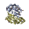
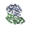
Reverse transcriptase domain of group II intron maturase from Roseburia intestinalis in P21 space group
Components
Retron-type reverse transcriptase
Keywords
RNA BINDING PROTEIN / Group II intron
Function / homology
Function and homology information
RNA-directed DNA polymerase / RNA-directed DNA polymerase activity / defense response to virus / RNA binding / metal ion binding Similarity search - Function
Group II intron, maturase-specific / Group II intron reverse transcriptase/maturase / Group II intron, maturase-specific domain / RNA-directed DNA polymerase (reverse transcriptase), msDNA / : / Reverse transcriptase (RNA-dependent DNA polymerase) / Reverse transcriptase domain / Reverse transcriptase (RT) catalytic domain profile. / DNA/RNA polymerase superfamily Similarity search - Domain/homology
In the structure databanks used in Yorodumi, some data are registered as the other names, "COVID-19 virus" and "2019-nCoV". Here are the details of the virus and the list of structure data.
Jan 31, 2019. EMDB accession codes are about to change! (news from PDBe EMDB page)
EMDB accession codes are about to change! (news from PDBe EMDB page)
The allocation of 4 digits for EMDB accession codes will soon come to an end. Whilst these codes will remain in use, new EMDB accession codes will include an additional digit and will expand incrementally as the available range of codes is exhausted. The current 4-digit format prefixed with “EMD-” (i.e. EMD-XXXX) will advance to a 5-digit format (i.e. EMD-XXXXX), and so on. It is currently estimated that the 4-digit codes will be depleted around Spring 2019, at which point the 5-digit format will come into force.
The EM Navigator/Yorodumi systems omit the EMD- prefix.
Related info.:Q: What is EMD? / ID/Accession-code notation in Yorodumi/EM Navigator
Yorodumi is a browser for structure data from EMDB, PDB, SASBDB, etc.
This page is also the successor to EM Navigator detail page, and also detail information page/front-end page for Omokage search.
The word "yorodu" (or yorozu) is an old Japanese word meaning "ten thousand". "mi" (miru) is to see.
Related info.:EMDB / PDB / SASBDB / Comparison of 3 databanks / Yorodumi Search / Aug 31, 2016. New EM Navigator & Yorodumi / Yorodumi Papers / Jmol/JSmol / Function and homology information / Changes in new EM Navigator and Yorodumi
 Movie
Movie Controller

 Yorodumi
 Open data
 Basic information
 Components
 Keywords
 Function and homology information
 Roseburia intestinalis XB6B4 (bacteria)
 X-RAY DIFFRACTION /
  Authors
 United States, 1items
  Citation
 Structure visualization
 Downloads & links
 Other downloads
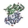


 PDBj
PDBj


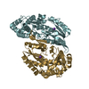
 Assembly


 Mass: 94.971 Da / Num. of mol.: 2 / Source method: obtained synthetically / Formula: PO4
 Mass: 39.098 Da / Num. of mol.: 2 / Source method: obtained synthetically / Formula: K
 Type: peptide linking / Mass: 75.067 Da / Num. of mol.: 3 / Source method: obtained synthetically / Formula: C2H5NO2
 Mass: 118.088 Da / Num. of mol.: 1 / Source method: obtained synthetically / Formula: C4H6O4
 Mass: 118.174 Da / Num. of mol.: 1 / Source method: obtained synthetically / Formula: C6H14O2 / Comment: precipitant*YM
 Sample preparation
 Processing