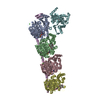
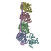
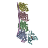
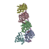
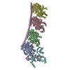
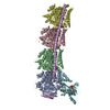
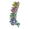
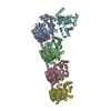
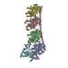
Entry Database : PDB / ID : 5h7oTitle Crystal structure of DJ-101 in complex with tubulin protein Stathmin-4 Tubulin alpha-1B chain Tubulin beta-2B chain Tubulin tyrosine ligase Keywords / / / / Function / homology Function Domain/homology Component
/ / / / / / / / / / / / / / / / / / / / / / / / / / / / / / / / / / / / / / / / / / / / / / / / / / / / / / / / / / / / / / / / / / / / / / / / / / / / / / / / / / / / / / / / / / / / / / / / / / / / / / / / / / / / / / / / / / / / / Biological species Sus scrofa (pig)Bos taurus (domestic cattle)Rattus norvegicus (Norway rat)Gallus gallus (chicken)Method / / Resolution : 2.8 Å Authors Arnst, K. / Wang, Y. / Hwang, D.-J. / Xue, Y. / Costello, T. / Hamilton, D. / Chen, Q. / Yang, J. / Park, F. / Dalton, J.T. ...Arnst, K. / Wang, Y. / Hwang, D.-J. / Xue, Y. / Costello, T. / Hamilton, D. / Chen, Q. / Yang, J. / Park, F. / Dalton, J.T. / Miller, D.D. / Li, W. Funding support Organization Grant number Country National Institutes of Health/National Cancer Institute (NIH/NCI) R01CA148706
Journal : Cancer Res. / Year : 2018Title : A Potent, Metabolically Stable Tubulin Inhibitor Targets the Colchicine Binding Site and Overcomes Taxane Resistance.Authors : Arnst, K.E. / Wang, Y. / Hwang, D.J. / Xue, Y. / Costello, T. / Hamilton, D. / Chen, Q. / Yang, J. / Park, F. / Dalton, J.T. / Miller, D.D. / Li, W. History Deposition Nov 20, 2016 Deposition site / Processing site Revision 1.0 Dec 13, 2017 Provider / Type Revision 1.1 Jan 17, 2018 Group / Database references / Category / diffrn_sourceItem _citation.journal_volume / _citation.page_first ... _citation.journal_volume / _citation.page_first / _citation.page_last / _citation.title / _citation.year / _diffrn_source.pdbx_synchrotron_beamline / _diffrn_source.pdbx_synchrotron_site Revision 1.2 Dec 4, 2019 Group / Category / Item Revision 1.3 Oct 12, 2022 Group Database references / Derived calculations ... Database references / Derived calculations / Other / Refinement description Category database_2 / pdbx_database_status ... database_2 / pdbx_database_status / pdbx_struct_conn_angle / refine_hist / struct_conn Item _database_2.pdbx_DOI / _database_2.pdbx_database_accession ... _database_2.pdbx_DOI / _database_2.pdbx_database_accession / _pdbx_database_status.deposit_site / _pdbx_struct_conn_angle.ptnr1_auth_asym_id / _pdbx_struct_conn_angle.ptnr1_auth_comp_id / _pdbx_struct_conn_angle.ptnr1_auth_seq_id / _pdbx_struct_conn_angle.ptnr1_label_asym_id / _pdbx_struct_conn_angle.ptnr1_label_atom_id / _pdbx_struct_conn_angle.ptnr1_label_comp_id / _pdbx_struct_conn_angle.ptnr1_label_seq_id / _pdbx_struct_conn_angle.ptnr2_auth_asym_id / _pdbx_struct_conn_angle.ptnr2_auth_comp_id / _pdbx_struct_conn_angle.ptnr2_auth_seq_id / _pdbx_struct_conn_angle.ptnr2_label_asym_id / _pdbx_struct_conn_angle.ptnr2_label_atom_id / _pdbx_struct_conn_angle.ptnr2_label_comp_id / _pdbx_struct_conn_angle.ptnr3_auth_asym_id / _pdbx_struct_conn_angle.ptnr3_auth_comp_id / _pdbx_struct_conn_angle.ptnr3_auth_seq_id / _pdbx_struct_conn_angle.ptnr3_label_asym_id / _pdbx_struct_conn_angle.ptnr3_label_atom_id / _pdbx_struct_conn_angle.ptnr3_label_comp_id / _pdbx_struct_conn_angle.ptnr3_label_seq_id / _pdbx_struct_conn_angle.value / _refine_hist.d_res_low / _struct_conn.pdbx_dist_value / _struct_conn.ptnr1_auth_asym_id / _struct_conn.ptnr1_auth_comp_id / _struct_conn.ptnr1_auth_seq_id / _struct_conn.ptnr1_label_asym_id / _struct_conn.ptnr1_label_atom_id / _struct_conn.ptnr1_label_comp_id / _struct_conn.ptnr1_label_seq_id / _struct_conn.ptnr2_auth_asym_id / _struct_conn.ptnr2_auth_comp_id / _struct_conn.ptnr2_auth_seq_id / _struct_conn.ptnr2_label_asym_id / _struct_conn.ptnr2_label_atom_id / _struct_conn.ptnr2_label_comp_id Revision 1.4 May 22, 2024 Group / Category / chem_comp_bond
Show all Show less
 Movie
Movie Controller


 Open data
 Basic information
 Components
 Keywords
 Function and homology information

 X-RAY DIFFRACTION /
  Authors
 United States, 1items
  Citation
 Structure visualization
 Downloads & links
 Other downloads
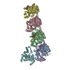


 PDBj
PDBj


 Assembly
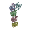


 Mass: 523.180 Da / Num. of mol.: 3 / Source method: obtained synthetically / Formula: C10H16N5O14P3 / Comment: GTP, energy-carrying molecule*YM
 Mass: 24.305 Da / Num. of mol.: 4 / Source method: obtained synthetically / Formula: Mg
 Mass: 40.078 Da / Num. of mol.: 2 / Source method: obtained synthetically / Formula: Ca
 Mass: 92.094 Da / Num. of mol.: 1 / Source method: obtained synthetically / Formula: C3H8O3
 Type: RNA linking / Mass: 443.201 Da / Num. of mol.: 1 / Source method: obtained synthetically / Formula: C10H15N5O11P2 / Comment: GDP, energy-carrying molecule*YM
 Mass: 195.237 Da / Num. of mol.: 1 / Source method: obtained synthetically / Formula: C6H13NO4S / Comment: pH buffer*YM
 Mass: 400.430 Da / Num. of mol.: 2 / Source method: obtained synthetically / Formula: C23H20N4O3
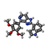
 Mass: 505.208 Da / Num. of mol.: 1 / Source method: obtained synthetically / Formula: C11H18N5O12P3 / Comment: AMP-PCP, energy-carrying molecule analogue*YM
 Sample preparation
 / Beamline: BL19U1 / Wavelength: 0.97853 Å
 Processing