 Movie
Movie Controller


+ Open data

- Basic information
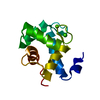
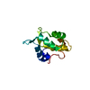
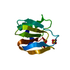
| Entry | Database: PDB / ID: 5h0p | ||||||
|---|---|---|---|---|---|---|---|
| Title | Crystal structure of EF-hand protein mutant | ||||||
 Components Components | EF-hand domain-containing protein D2 | ||||||
 Keywords Keywords | METAL BINDING PROTEIN / EF-hand / Phosphorylation | ||||||
| Function / homology |  Function and homology information Function and homology informationRHOD GTPase cycle / cadherin binding / membrane raft / calcium ion binding Similarity search - Function | ||||||
| Biological species |  Homo sapiens (human) Homo sapiens (human) | ||||||
| Method |  X-RAY DIFFRACTION / SYNCHROTRON / MOLECULAR REPLACEMENT / Resolution: 1.862 Å X-RAY DIFFRACTION / SYNCHROTRON / MOLECULAR REPLACEMENT / Resolution: 1.862 Å | ||||||
 Authors Authors | Park, K.R. / An, J.Y. / Kang, J.Y. / Lee, J.G. / Youn, H.S. / Lee, Y. / Mun, S.A. / Jun, C.D. / Song, W.K. / Eom, S.H. | ||||||
 Citation Citation | Journal: Biochem. Biophys. Res. Commun. / Year: 2017 Title: Structural mechanism underlying regulation of human EFhd2/Swiprosin-1 actin-bundling activity by Ser183 phosphorylation. Authors: Park, K.R. / An, J.Y. / Kang, J.Y. / Lee, J.G. / Lee, Y. / Mun, S.A. / Jun, C.D. / Song, W.K. / Eom, S.H. | ||||||
| History |
|
- Structure visualization
| Structure viewer | Molecule: MolmilJmol/JSmol |
|---|
- Downloads & links
-Download
| PDBx/mmCIF format | 5h0p.cif.gz | 58.2 KB | Display | PDBx/mmCIF format |
|---|---|---|---|---|
| PDB format | pdb5h0p.ent.gz | 40.4 KB | Display | PDB format |
| PDBx/mmJSON format | 5h0p.json.gz | Tree view | PDBx/mmJSON format | |
| Others |  Other downloads Other downloads |
-Validation report
| Summary document | 5h0p_validation.pdf.gz | 416.1 KB | Display | wwPDB validaton report |
|---|---|---|---|---|
| Full document | 5h0p_full_validation.pdf.gz | 416 KB | Display | |
| Data in XML | 5h0p_validation.xml.gz | 6.2 KB | Display | |
| Data in CIF | 5h0p_validation.cif.gz | 7.5 KB | Display | |
| Arichive directory | https://data.pdbj.org/pub/pdb/validation_reports/h0/5h0pftp://data.pdbj.org/pub/pdb/validation_reports/h0/5h0p | HTTPS FTP |
-Related structure data
| Related structure data |  5i2lS S: Starting model for refinement |
|---|---|
| Similar structure data |


-Links
 PDBj
PDBj- Assembly
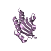
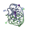
| Deposited unit | 
| ||||||||
|---|---|---|---|---|---|---|---|---|---|
| 1 |
| ||||||||
| Unit cell |
|
-Components
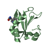
| #1: Protein | Mass: 13751.778 Da / Num. of mol.: 1 / Fragment: UNP RESIDUES 70-184 / Mutation: S183E Source method: isolated from a genetically manipulated source Source: (gene. exp.) Homo sapiens (human) / Gene: EFHD2, SWS1 / Plasmid: modified pET28a / Production host:  | ||
|---|---|---|---|
| #2: Chemical |   Mass: 40.078 Da / Num. of mol.: 2 / Source method: obtained synthetically / Formula: Ca Mass: 40.078 Da / Num. of mol.: 2 / Source method: obtained synthetically / Formula: Ca#3: Water | ChemComp-HOH / |  Mass: 18.015 Da / Num. of mol.: 39 / Source method: isolated from a natural source / Formula: H2O Mass: 18.015 Da / Num. of mol.: 39 / Source method: isolated from a natural source / Formula: H2O |
-Experimental details
-Experiment
| Experiment | Method: X-RAY DIFFRACTION / Number of used crystals: 1 |
|---|
- Sample preparation
| Crystal | Density Matthews: 1.83 Å3/Da / Density % sol: 32.71 % |
|---|---|
| Crystal grow | Temperature: 290 K / Method: vapor diffusion, hanging drop / pH: 8.5 Details: 0.2 M trimethlyamine-N-oxide dihydrate, 0.1 M Tris-HCl (pH 8.5), 20% (w/v) PEG 2000 MME |
-Data collection
| Diffraction | Mean temperature: 80 K |
|---|---|
| Diffraction source | Source: SYNCHROTRON / Site: PAL/PLS  / Beamline: 5C (4A) / Wavelength: 0.9897 Å / Beamline: 5C (4A) / Wavelength: 0.9897 Å |
| Detector | Type: ADSC QUANTUM 315r / Detector: CCD / Date: Oct 23, 2013 |
| Radiation | Protocol: SINGLE WAVELENGTH / Monochromatic (M) / Laue (L): M / Scattering type: x-ray |
| Radiation wavelength | Wavelength: 0.9897 Å / Relative weight: 1 |
| Reflection | Resolution: 1.86→50 Å / Num. obs: 8596 / % possible obs: 96.9 % / Redundancy: 13.2 % / Net I/σ(I): 22.7 |
| Reflection shell | Resolution: 1.86→1.89 Å / Redundancy: 13.2 % / Rmerge(I) obs: 0.315 / Mean I/σ(I) obs: 6.6 / % possible all: 98.4 |
- Processing
| Software |
| |||||||||||||||||||||||||||||||||||||||||||||||||||||||||||||||||||||||||||||||||||||||||||||||||||||||||||||||||||||||||||||
|---|---|---|---|---|---|---|---|---|---|---|---|---|---|---|---|---|---|---|---|---|---|---|---|---|---|---|---|---|---|---|---|---|---|---|---|---|---|---|---|---|---|---|---|---|---|---|---|---|---|---|---|---|---|---|---|---|---|---|---|---|---|---|---|---|---|---|---|---|---|---|---|---|---|---|---|---|---|---|---|---|---|---|---|---|---|---|---|---|---|---|---|---|---|---|---|---|---|---|---|---|---|---|---|---|---|---|---|---|---|---|---|---|---|---|---|---|---|---|---|---|---|---|---|---|---|---|
| Refinement | Method to determine structure: MOLECULAR REPLACEMENT Starting model: 5I2L Resolution: 1.862→29.796 Å / SU ML: 0.19 / Cross valid method: NONE / σ(F): 1.37 / Phase error: 27.37
| |||||||||||||||||||||||||||||||||||||||||||||||||||||||||||||||||||||||||||||||||||||||||||||||||||||||||||||||||||||||||||||
| Solvent computation | Shrinkage radii: 0.9 Å / VDW probe radii: 1.11 Å | |||||||||||||||||||||||||||||||||||||||||||||||||||||||||||||||||||||||||||||||||||||||||||||||||||||||||||||||||||||||||||||
| Refinement step | Cycle: LAST / Resolution: 1.862→29.796 Å
| |||||||||||||||||||||||||||||||||||||||||||||||||||||||||||||||||||||||||||||||||||||||||||||||||||||||||||||||||||||||||||||
| Refine LS restraints |
| |||||||||||||||||||||||||||||||||||||||||||||||||||||||||||||||||||||||||||||||||||||||||||||||||||||||||||||||||||||||||||||
| LS refinement shell |
| |||||||||||||||||||||||||||||||||||||||||||||||||||||||||||||||||||||||||||||||||||||||||||||||||||||||||||||||||||||||||||||
| Refinement TLS params. | Method: refined / Refine-ID: X-RAY DIFFRACTION
| |||||||||||||||||||||||||||||||||||||||||||||||||||||||||||||||||||||||||||||||||||||||||||||||||||||||||||||||||||||||||||||
| Refinement TLS group |
|
