 Movie
Movie Controller

[English] 日本語
 Yorodumi
- PDB-5guj: Crystal structure of the Bacillus subtilis DnaG RNA Polymerase Do... -
+ Open data

- Basic information
| Entry | Database: PDB / ID: 5guj | ||||||
|---|---|---|---|---|---|---|---|
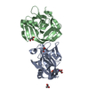
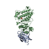
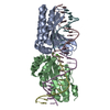
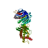
| Title | Crystal structure of the Bacillus subtilis DnaG RNA Polymerase Domain, natural degradation of full length DnaG | ||||||
 Components Components | DNA primase | ||||||
 Keywords Keywords | TRANSFERASE | ||||||
| Function / homology |  Function and homology information Function and homology informationDNA primase DnaG / primosome complex / DNA replication, synthesis of primer / DNA helicase activity / DNA-directed RNA polymerase complex / DNA-directed RNA polymerase activity / DNA binding / zinc ion binding / ATP binding / cytoplasm Similarity search - Function | ||||||
| Biological species |  | ||||||
| Method |  X-RAY DIFFRACTION / SYNCHROTRON / MOLECULAR REPLACEMENT / Resolution: 2.5 Å X-RAY DIFFRACTION / SYNCHROTRON / MOLECULAR REPLACEMENT / Resolution: 2.5 Å | ||||||
 Authors Authors | Zhou, Y. / Liu, Z. / Wang, G. | ||||||
 Citation Citation | Journal: Sci Rep / Year: 2017 Title: Structural Insight into the Specific DNA Template Binding to DnaG primase in Bacteria Authors: Zhou, Y. / Luo, H. / Liu, Z. / Yang, M. / Pang, X. / Sun, F. / Wang, G. | ||||||
| History |
|
- Structure visualization
| Structure viewer | Molecule: MolmilJmol/JSmol |
|---|
- Downloads & links
-Download
| PDBx/mmCIF format | 5guj.cif.gz | 80 KB | Display | PDBx/mmCIF format |
|---|---|---|---|---|
| PDB format | pdb5guj.ent.gz | 57.9 KB | Display | PDB format |
| PDBx/mmJSON format | 5guj.json.gz | Tree view | PDBx/mmJSON format | |
| Others |  Other downloads Other downloads |
-Validation report
| Arichive directory | https://data.pdbj.org/pub/pdb/validation_reports/gu/5gujftp://data.pdbj.org/pub/pdb/validation_reports/gu/5guj | HTTPS FTP |
|---|
-Related structure data
| Related structure data | 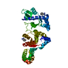 4e2kS  5gum S: Starting model for refinement |
|---|---|
| Similar structure data |


-Links
 PDBj
PDBj

- Assembly
| Deposited unit | 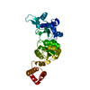
| ||||||||
|---|---|---|---|---|---|---|---|---|---|
| 1 |
| ||||||||
| Unit cell |
|
-Components
| #1: Protein | Mass: 36987.234 Da / Num. of mol.: 1 / Fragment: UNP residues 112-435 Source method: isolated from a genetically manipulated source Source: (gene. exp.) Strain: 168 / Gene: dnaG, dnaE, BSU25210 / Plasmid: pGEX-6p-1 / Production host: References: UniProt: P05096, Transferases; Transferring phosphorus-containing groups; Nucleotidyltransferases |
|---|---|
| #2: Water | ChemComp-HOH /  Mass: 18.015 Da / Num. of mol.: 76 / Source method: isolated from a natural source / Formula: H2O Mass: 18.015 Da / Num. of mol.: 76 / Source method: isolated from a natural source / Formula: H2O |
-Experimental details
-Experiment
| Experiment | Method: X-RAY DIFFRACTION / Number of used crystals: 1 |
|---|
- Sample preparation
| Crystal | Density Matthews: 2.62 Å3/Da / Density % sol: 52.96 % |
|---|---|
| Crystal grow | Temperature: 291 K / Method: vapor diffusion, hanging drop / pH: 8.5 Details: 0.2 M Sodium citrate tribasic dehydrate, 0.1M Tris hydrochloride (pH 8.5) and 30% (w/v) polyethylene glycol 400 |
-Data collection
| Diffraction | Mean temperature: 100 K |
|---|---|
| Diffraction source | Source: SYNCHROTRON / Site: SSRF  / Beamline: BL17U / Wavelength: 0.97916 Å / Beamline: BL17U / Wavelength: 0.97916 Å |
| Detector | Type: ADSC QUANTUM 315r / Detector: CCD / Date: May 5, 2016 |
| Radiation | Protocol: SINGLE WAVELENGTH / Monochromatic (M) / Laue (L): M / Scattering type: x-ray |
| Radiation wavelength | Wavelength: 0.97916 Å / Relative weight: 1 |
| Reflection | Resolution: 2.5→29.278 Å / Num. obs: 13480 / % possible obs: 100 % / Redundancy: 13.3 % / Rmerge(I) obs: 0.123 / Net I/σ(I): 20.7 |
| Reflection shell | Resolution: 2.5→2.54 Å / Redundancy: 13.5 % / Rmerge(I) obs: 0.429 / Mean I/σ(I) obs: 6.5 / % possible all: 100 |
- Processing
| Software |
| |||||||||||||||||||||||||||||||||||
|---|---|---|---|---|---|---|---|---|---|---|---|---|---|---|---|---|---|---|---|---|---|---|---|---|---|---|---|---|---|---|---|---|---|---|---|---|
| Refinement | Method to determine structure: MOLECULAR REPLACEMENT Starting model: 4e2k Resolution: 2.5→29.278 Å / SU ML: 0.23 / Cross valid method: THROUGHOUT / σ(F): 1.34 / Phase error: 25
| |||||||||||||||||||||||||||||||||||
| Solvent computation | Shrinkage radii: 0.9 Å / VDW probe radii: 1.11 Å | |||||||||||||||||||||||||||||||||||
| Refinement step | Cycle: LAST / Resolution: 2.5→29.278 Å
| |||||||||||||||||||||||||||||||||||
| Refine LS restraints |
| |||||||||||||||||||||||||||||||||||
| LS refinement shell |
|
