 Movie
Movie Controller

[English] 日本語
 Yorodumi
- PDB-5wtb: Complex Structure of Staphylococcus aureus SdrE with human comple... -
+ Open data

- Basic information
| Entry | Database: PDB / ID: 5wtb | ||||||
|---|---|---|---|---|---|---|---|
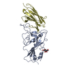
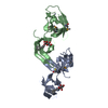
| Title | Complex Structure of Staphylococcus aureus SdrE with human complement factor H | ||||||
 Components Components |
| ||||||
 Keywords Keywords | CELL ADHESION / Staphylococcus aureus / Sdr family / complement factor H / complement evasion | ||||||
| Function / homology |  Function and homology information Function and homology informationregulation of complement activation, alternative pathway / regulation of complement-dependent cytotoxicity / regulation of complement activation / complement component C3b binding / central nervous system myelination / symbiont cell surface / serine-type endopeptidase complex / heparan sulfate proteoglycan binding / complement activation, alternative pathway / complement activation ...regulation of complement activation, alternative pathway / regulation of complement-dependent cytotoxicity / regulation of complement activation / complement component C3b binding / central nervous system myelination / symbiont cell surface / serine-type endopeptidase complex / heparan sulfate proteoglycan binding / complement activation, alternative pathway / complement activation / Regulation of Complement cascade / heparin binding / blood microparticle / cell adhesion / inflammatory response / proteolysis / : / extracellular exosome / extracellular region / identical protein binding Similarity search - Function | ||||||
| Biological species |   Staphylococcus aureus (bacteria) Staphylococcus aureus (bacteria) Homo sapiens (human) Homo sapiens (human) | ||||||
| Method |  X-RAY DIFFRACTION / SYNCHROTRON / MOLECULAR REPLACEMENT / Resolution: 3.3 Å X-RAY DIFFRACTION / SYNCHROTRON / MOLECULAR REPLACEMENT / Resolution: 3.3 Å | ||||||
 Authors Authors | Wu, M. / Zhang, Y. / Hang, T. / Wang, C. / Yang, Y. / Zang, J. / Zhang, M. / Zhang, X. | ||||||
 Citation Citation | Journal: Biochem. J. / Year: 2017 Title: Staphylococcus aureus SdrE captures complement factor H's C-terminus via a novel 'close, dock, lock and latch' mechanism for complement evasion Authors: Zhang, Y. / Wu, M. / Hang, T. / Wang, C. / Yang, Y. / Pan, W. / Zang, J. / Zhang, M. / Zhang, X. | ||||||
| History |
|
- Structure visualization
| Structure viewer | Molecule: MolmilJmol/JSmol |
|---|
- Downloads & links
-Download
| PDBx/mmCIF format | 5wtb.cif.gz | 266.9 KB | Display | PDBx/mmCIF format |
|---|---|---|---|---|
| PDB format | pdb5wtb.ent.gz | 215 KB | Display | PDB format |
| PDBx/mmJSON format | 5wtb.json.gz | Tree view | PDBx/mmJSON format | |
| Others |  Other downloads Other downloads |
-Validation report
| Arichive directory | https://data.pdbj.org/pub/pdb/validation_reports/wt/5wtbftp://data.pdbj.org/pub/pdb/validation_reports/wt/5wtb | HTTPS FTP |
|---|
-Related structure data
| Related structure data |  5wtaC  1r17S C: citing same article ( S: Starting model for refinement |
|---|---|
| Similar structure data |


-Links
 PDBj
PDBj

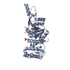
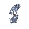
- Assembly
| Deposited unit | 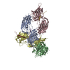
| ||||||||
|---|---|---|---|---|---|---|---|---|---|
| 1 | 
| ||||||||
| 2 | 
| ||||||||
| 3 | 
| ||||||||
| 4 | 
| ||||||||
| Unit cell |
|
-Components
| #1: Protein | Mass: 37694.152 Da / Num. of mol.: 4 / Fragment: ligand binding A-domain, UNP residues 270-599 / Mutation: I489M Source method: isolated from a genetically manipulated source Source: (gene. exp.) Staphylococcus aureus (strain Mu50 / ATCC 700699) (bacteria)Strain: Mu50 / ATCC 700699 / Gene: sdrE, SAV0563 / Plasmid: pET22b / Production host: #2: Protein/peptide | Mass: 2511.836 Da / Num. of mol.: 4 / Fragment: UNP residues 1206-1226 / Source method: obtained synthetically / Source: (synth.) Homo sapiens (human) / References: UniProt: P08603 |
|---|
-Experimental details
-Experiment
| Experiment | Method: X-RAY DIFFRACTION / Number of used crystals: 1 |
|---|
- Sample preparation
| Crystal | Density Matthews: 3.46 Å3/Da / Density % sol: 64.41 % |
|---|---|
| Crystal grow | Temperature: 298 K / Method: vapor diffusion, sitting drop Details: 0.1M HEPES sodium pH 7.5, 0.2M Magnesium chloride, 30% PEG 400 (v/v) |
-Data collection
| Diffraction | Mean temperature: 100 K |
|---|---|
| Diffraction source | Source: SYNCHROTRON / Site: SSRF  / Beamline: BL18U1 / Wavelength: 0.9779 Å / Beamline: BL18U1 / Wavelength: 0.9779 Å |
| Detector | Type: DECTRIS PILATUS3 6M / Detector: PIXEL / Date: Mar 21, 2016 |
| Radiation | Protocol: SINGLE WAVELENGTH / Monochromatic (M) / Laue (L): M / Scattering type: x-ray |
| Radiation wavelength | Wavelength: 0.9779 Å / Relative weight: 1 |
| Reflection | Resolution: 3.3→49.79 Å / Num. obs: 31291 / % possible obs: 96.1 % / Redundancy: 2.8 % / Rsym value: 0.1 / Net I/σ(I): 7 |
| Reflection shell | Resolution: 3.3→3.48 Å / Redundancy: 2.8 % / Mean I/σ(I) obs: 1.9 / Num. measured obs: 4613 / Rsym value: 0.56 / % possible all: 97.9 |
- Processing
| Software |
| ||||||||||||||||||||||||||||||||||||||||||||||||||||||||||||||||||||||||||||||||||||||||||||||||||||||||||||||||||||||||||||||||||||||||||||||||||||||||||||||||||||||||||||||||||||||
|---|---|---|---|---|---|---|---|---|---|---|---|---|---|---|---|---|---|---|---|---|---|---|---|---|---|---|---|---|---|---|---|---|---|---|---|---|---|---|---|---|---|---|---|---|---|---|---|---|---|---|---|---|---|---|---|---|---|---|---|---|---|---|---|---|---|---|---|---|---|---|---|---|---|---|---|---|---|---|---|---|---|---|---|---|---|---|---|---|---|---|---|---|---|---|---|---|---|---|---|---|---|---|---|---|---|---|---|---|---|---|---|---|---|---|---|---|---|---|---|---|---|---|---|---|---|---|---|---|---|---|---|---|---|---|---|---|---|---|---|---|---|---|---|---|---|---|---|---|---|---|---|---|---|---|---|---|---|---|---|---|---|---|---|---|---|---|---|---|---|---|---|---|---|---|---|---|---|---|---|---|---|---|---|
| Refinement | Method to determine structure: MOLECULAR REPLACEMENT Starting model: 1R17 Resolution: 3.3→49.79 Å / Cor.coef. Fo:Fc: 0.925 / Cor.coef. Fo:Fc free: 0.886 / Cross valid method: THROUGHOUT / ESU R Free: 0.568 / Details: HYDROGENS HAVE BEEN ADDED IN THE RIDING POSITIONS
| ||||||||||||||||||||||||||||||||||||||||||||||||||||||||||||||||||||||||||||||||||||||||||||||||||||||||||||||||||||||||||||||||||||||||||||||||||||||||||||||||||||||||||||||||||||||
| Solvent computation | Ion probe radii: 0.8 Å / Shrinkage radii: 0.8 Å / VDW probe radii: 1.2 Å | ||||||||||||||||||||||||||||||||||||||||||||||||||||||||||||||||||||||||||||||||||||||||||||||||||||||||||||||||||||||||||||||||||||||||||||||||||||||||||||||||||||||||||||||||||||||
| Displacement parameters | Biso mean: 89.516 Å2
| ||||||||||||||||||||||||||||||||||||||||||||||||||||||||||||||||||||||||||||||||||||||||||||||||||||||||||||||||||||||||||||||||||||||||||||||||||||||||||||||||||||||||||||||||||||||
| Refinement step | Cycle: 1 / Resolution: 3.3→49.79 Å
| ||||||||||||||||||||||||||||||||||||||||||||||||||||||||||||||||||||||||||||||||||||||||||||||||||||||||||||||||||||||||||||||||||||||||||||||||||||||||||||||||||||||||||||||||||||||
| Refine LS restraints |
|