 Movie
Movie Controller

[English] 日本語
 Yorodumi
- PDB-4aag: Crystal structure of the mutant D75N I-CreI in complex with its w... -
+ Open data

- Basic information
| Entry | Database: PDB / ID: 4aag | ||||||
|---|---|---|---|---|---|---|---|
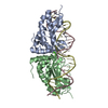
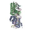
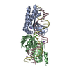
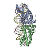
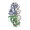
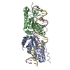
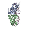
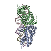
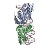
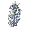
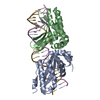
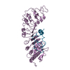
| Title | Crystal structure of the mutant D75N I-CreI in complex with its wild- type target in presence of Ca at the active site (The four central bases, 2NN region, are composed by GTAC from 5' to 3') | ||||||
 Components Components |
| ||||||
 Keywords Keywords | HYDROLASE/DNA / HYDROLASE-DNA COMPLEX / GENE TARGETING / PROTEIN-DNA INTERACTION / HOMING ENDONUCLEASES | ||||||
| Function / homology |  Function and homology information Function and homology informationintron homing / chloroplast / endonuclease activity / Hydrolases; Acting on ester bonds / hydrolase activity / metal ion binding / identical protein binding Similarity search - Function | ||||||
| Biological species |   CHLAMYDOMONAS REINHARDTII (plant) CHLAMYDOMONAS REINHARDTII (plant)SYNTHETIC CONSTRUCT (others) | ||||||
| Method |  X-RAY DIFFRACTION / SYNCHROTRON / MOLECULAR REPLACEMENT / Resolution: 2.8 Å X-RAY DIFFRACTION / SYNCHROTRON / MOLECULAR REPLACEMENT / Resolution: 2.8 Å | ||||||
 Authors Authors | Molina, R. / Redondo, P. / Stella, S. / Marenchino, M. / D'Abramo, M. / Gervasio, F. / Epinat, J.C. / Valton, J. / Grizot, S. / Duchateau, P. ...Molina, R. / Redondo, P. / Stella, S. / Marenchino, M. / D'Abramo, M. / Gervasio, F. / Epinat, J.C. / Valton, J. / Grizot, S. / Duchateau, P. / Prieto, J. / Montoya, G. | ||||||
 Citation Citation | Journal: Nucleic Acids Res. / Year: 2012 Title: Non-Specific Protein-DNA Interactions Control I-Crei Target Binding and Cleavage. Authors: Molina, R. / Redondo, P. / Stella, S. / Marenchino, M. / D'Abramo, M. / Gervasio, F.L. / Charles Epinat, J. / Valton, J. / Grizot, S. / Duchateau, P. / Prieto, J. / Montoya, G. | ||||||
| History |
|
- Structure visualization
| Structure viewer | Molecule: MolmilJmol/JSmol |
|---|
- Downloads & links
-Download
| PDBx/mmCIF format | 4aag.cif.gz | 194.4 KB | Display | PDBx/mmCIF format |
|---|---|---|---|---|
| PDB format | pdb4aag.ent.gz | 149.8 KB | Display | PDB format |
| PDBx/mmJSON format | 4aag.json.gz | Tree view | PDBx/mmJSON format | |
| Others |  Other downloads Other downloads |
-Validation report
| Arichive directory | https://data.pdbj.org/pub/pdb/validation_reports/aa/4aagftp://data.pdbj.org/pub/pdb/validation_reports/aa/4aag | HTTPS FTP |
|---|
-Related structure data
| Related structure data |  4aabC  4aadC  4aaeC  4aafC  2xe0S C: citing same article ( S: Starting model for refinement |
|---|---|
| Similar structure data |


-Links
 PDBj
PDBj


- Assembly
| Deposited unit | 
| ||||||||
|---|---|---|---|---|---|---|---|---|---|
| 1 |
| ||||||||
| Unit cell |
|
-Components
| #1: Protein | Mass: 17515.137 Da / Num. of mol.: 2 / Mutation: YES Source method: isolated from a genetically manipulated source Source: (gene. exp.) CHLAMYDOMONAS REINHARDTII (plant) / Production host:  References: UniProt: P05725, Hydrolases; Acting on ester bonds #2: DNA chain | Mass: 7368.778 Da / Num. of mol.: 2 / Source method: obtained synthetically / Source: (synth.) SYNTHETIC CONSTRUCT (others) #3: Chemical |   Mass: 40.078 Da / Num. of mol.: 2 / Source method: obtained synthetically / Formula: Ca Mass: 40.078 Da / Num. of mol.: 2 / Source method: obtained synthetically / Formula: Ca#4: Water | ChemComp-HOH / |  Mass: 18.015 Da / Num. of mol.: 40 / Source method: isolated from a natural source / Formula: H2O Mass: 18.015 Da / Num. of mol.: 40 / Source method: isolated from a natural source / Formula: H2OCompound details | ENGINEERED | |
|---|
-Experimental details
-Experiment
| Experiment | Method: X-RAY DIFFRACTION / Number of used crystals: 1 |
|---|
- Sample preparation
| Crystal | Density Matthews: 2.9 Å3/Da / Density % sol: 62 % / Description: NONE |
|---|---|
| Crystal grow | pH: 7.5 / Details: 30% PEG400, 0.1M HEPES PH 7.5, 0.1M NACL |
-Data collection
| Diffraction | Mean temperature: 100 K |
|---|---|
| Diffraction source | Source: SYNCHROTRON / Site: SLS  / Beamline: X06SA / Wavelength: 1 / Beamline: X06SA / Wavelength: 1 |
| Detector | Type: DECTRIS PILATUS 6M / Detector: PIXEL |
| Radiation | Protocol: SINGLE WAVELENGTH / Monochromatic (M) / Laue (L): M / Scattering type: x-ray |
| Radiation wavelength | Wavelength: 1 Å / Relative weight: 1 |
| Reflection | Resolution: 2.8→47.47 Å / Num. obs: 15005 / % possible obs: 99.8 % / Observed criterion σ(I): 1.5 / Redundancy: 4.2 % / Biso Wilson estimate: 57.29 Å2 / Rmerge(I) obs: 0.09 / Net I/σ(I): 5.3 |
| Reflection shell | Resolution: 2.8→2.95 Å / Redundancy: 3.9 % / Rmerge(I) obs: 0.48 / Mean I/σ(I) obs: 1.5 / % possible all: 99.7 |
- Processing
| Software |
| ||||||||||||||||||||||||||||||||||||||||||||||||||||||||||||||||||||||||||||||||||||
|---|---|---|---|---|---|---|---|---|---|---|---|---|---|---|---|---|---|---|---|---|---|---|---|---|---|---|---|---|---|---|---|---|---|---|---|---|---|---|---|---|---|---|---|---|---|---|---|---|---|---|---|---|---|---|---|---|---|---|---|---|---|---|---|---|---|---|---|---|---|---|---|---|---|---|---|---|---|---|---|---|---|---|---|---|---|
| Refinement | Method to determine structure: MOLECULAR REPLACEMENT Starting model: PDB ENTRY 2XE0 Resolution: 2.8→45.743 Å / SU ML: 0.77 / σ(F): 1.34 / Phase error: 26.51 / Stereochemistry target values: ML
| ||||||||||||||||||||||||||||||||||||||||||||||||||||||||||||||||||||||||||||||||||||
| Solvent computation | Shrinkage radii: 0.72 Å / VDW probe radii: 1 Å / Solvent model: FLAT BULK SOLVENT MODEL / Bsol: 55.086 Å2 / ksol: 0.336 e/Å3 | ||||||||||||||||||||||||||||||||||||||||||||||||||||||||||||||||||||||||||||||||||||
| Displacement parameters |
| ||||||||||||||||||||||||||||||||||||||||||||||||||||||||||||||||||||||||||||||||||||
| Refinement step | Cycle: LAST / Resolution: 2.8→45.743 Å
| ||||||||||||||||||||||||||||||||||||||||||||||||||||||||||||||||||||||||||||||||||||
| Refine LS restraints |
| ||||||||||||||||||||||||||||||||||||||||||||||||||||||||||||||||||||||||||||||||||||
| LS refinement shell |
| ||||||||||||||||||||||||||||||||||||||||||||||||||||||||||||||||||||||||||||||||||||
| Refinement TLS params. | Method: refined / Origin x: 12.1361 Å / Origin y: -27.0071 Å / Origin z: -23.6519 Å
| ||||||||||||||||||||||||||||||||||||||||||||||||||||||||||||||||||||||||||||||||||||
| Refinement TLS group | Selection details: ALL |
