 Movie
Movie Controller

[English] 日本語
 Yorodumi
- PDB-5gmu: Crystal structure of chorismate mutase like domain of bifunctiona... -
+ Open data

- Basic information
| Entry | Database: PDB / ID: 5gmu | ||||||
|---|---|---|---|---|---|---|---|
| Title | Crystal structure of chorismate mutase like domain of bifunctional DAHP synthase of Bacillus subtilis in complex with Chlorogenic acid | ||||||
 Components Components | Protein AroA(G) | ||||||
 Keywords Keywords | ISOMERASE / Type II chorismate mutase / CML domain / Bifunctional DAHP synthase / Chlorogenic acid | ||||||
| Function / homology |  Function and homology information Function and homology informationchorismate mutase / chorismate mutase activity / 3-deoxy-7-phosphoheptulonate synthase / 3-deoxy-7-phosphoheptulonate synthase activity / aldehyde-lyase activity / chorismate biosynthetic process / aromatic amino acid biosynthetic process / amino acid biosynthetic process / cytoplasm Similarity search - Function | ||||||
| Biological species |  | ||||||
| Method |  X-RAY DIFFRACTION / MOLECULAR REPLACEMENT / Resolution: 1.8 Å X-RAY DIFFRACTION / MOLECULAR REPLACEMENT / Resolution: 1.8 Å | ||||||
 Authors Authors | Pratap, S. / Dev, A. / Sharma, V. / Yadav, R. / Narwal, M. / Tomar, S. / Kumar, P. | ||||||
 Citation Citation | Journal: Sci Rep / Year: 2017 Title: Structure of Chorismate Mutase-like Domain of DAHPS from Bacillus subtilis Complexed with Novel Inhibitor Reveals Conformational Plasticity of Active Site. Authors: Pratap, S. / Dev, A. / Kumar, V. / Yadav, R. / Narwal, M. / Tomar, S. / Kumar, P. | ||||||
| History |
|
- Structure visualization
| Structure viewer | Molecule: MolmilJmol/JSmol |
|---|
- Downloads & links
-Download
| PDBx/mmCIF format | 5gmu.cif.gz | 89.5 KB | Display | PDBx/mmCIF format |
|---|---|---|---|---|
| PDB format | pdb5gmu.ent.gz | 68.1 KB | Display | PDB format |
| PDBx/mmJSON format | 5gmu.json.gz | Tree view | PDBx/mmJSON format | |
| Others |  Other downloads Other downloads |
-Validation report
| Arichive directory | https://data.pdbj.org/pub/pdb/validation_reports/gm/5gmuftp://data.pdbj.org/pub/pdb/validation_reports/gm/5gmu | HTTPS FTP |
|---|
-Related structure data
| Related structure data |  5go2C  3nvtS C: citing same article ( S: Starting model for refinement |
|---|---|
| Similar structure data |


-Links
 PDBj
PDBj
- Assembly
| Deposited unit | 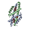
| ||||||||||||||||||
|---|---|---|---|---|---|---|---|---|---|---|---|---|---|---|---|---|---|---|---|
| 1 | 
| ||||||||||||||||||
| 2 | 
| ||||||||||||||||||
| Unit cell |
| ||||||||||||||||||
| Noncrystallographic symmetry (NCS) | NCS domain:
NCS domain segments: Component-ID: _ / Ens-ID: 1 / Beg auth comp-ID: THR / Beg label comp-ID: THR / End auth comp-ID: GLU / End label comp-ID: GLU / Refine code: _ / Auth seq-ID: 4 - 87 / Label seq-ID: 4 - 87
|
-Components
| #1: Protein | Mass: 10430.755 Da / Num. of mol.: 2 Fragment: Chorismate Mutase type II like domain, UNP residues 1-90 Source method: isolated from a genetically manipulated source Source: (gene. exp.) Strain: 168 / Gene: aroA, BSU29750 Production host: Strain (production host): BL21-Gold(DE3)pLysS AG / References: UniProt: P39912, chorismate mutase #2: Chemical |   Mass: 356.325 Da / Num. of mol.: 2 / Source method: obtained synthetically / Formula: C16H20O9 Mass: 356.325 Da / Num. of mol.: 2 / Source method: obtained synthetically / Formula: C16H20O9#3: Chemical | ChemComp-SO4 / |   Mass: 96.063 Da / Num. of mol.: 1 / Source method: obtained synthetically / Formula: SO4 Mass: 96.063 Da / Num. of mol.: 1 / Source method: obtained synthetically / Formula: SO4#4: Water | ChemComp-HOH / |  Mass: 18.015 Da / Num. of mol.: 116 / Source method: isolated from a natural source / Formula: H2O Mass: 18.015 Da / Num. of mol.: 116 / Source method: isolated from a natural source / Formula: H2O |
|---|
-Experimental details
-Experiment
| Experiment | Method: X-RAY DIFFRACTION / Number of used crystals: 1 |
|---|
- Sample preparation
| Crystal | Density Matthews: 2.18 Å3/Da / Density % sol: 43.65 % |
|---|---|
| Crystal grow | Temperature: 293 K / Method: vapor diffusion, hanging drop / pH: 5.6 Details: 0.2M Potassium sodium tartrate tetrahydrate, 0.1M Sodium citrate tribasic dihydrate pH 5.6, 2.0M Ammonium sulfate |
-Data collection
| Diffraction | Mean temperature: 100 K |
|---|---|
| Diffraction source | Source: ROTATING ANODE / Type: BRUKER AXS MICROSTAR-H / Wavelength: 1.54 Å |
| Detector | Type: MAR scanner 345 mm plate / Detector: IMAGE PLATE / Date: May 20, 2015 |
| Radiation | Protocol: SINGLE WAVELENGTH / Monochromatic (M) / Laue (L): M / Scattering type: x-ray |
| Radiation wavelength | Wavelength: 1.54 Å / Relative weight: 1 |
| Reflection | Resolution: 1.8→50 Å / Num. obs: 16372 / % possible obs: 98 % / Redundancy: 3.4 % / Rmerge(I) obs: 0.06 / Net I/σ(I): 24.58 |
| Reflection shell | Resolution: 1.8→1.83 Å / Redundancy: 2.7 % / Rmerge(I) obs: 0.266 / Mean I/σ(I) obs: 3.76 / CC1/2: 0.926 / % possible all: 88 |
- Processing
| Software |
| ||||||||||||||||||||||||||||||||||||||||||||||||||||||||||||||||||||||||||||||||||||||||||||||||||||||||||||||||||||||||||||||||||||||||||||||||||||||||||||||||||||||||||||||||||||||
|---|---|---|---|---|---|---|---|---|---|---|---|---|---|---|---|---|---|---|---|---|---|---|---|---|---|---|---|---|---|---|---|---|---|---|---|---|---|---|---|---|---|---|---|---|---|---|---|---|---|---|---|---|---|---|---|---|---|---|---|---|---|---|---|---|---|---|---|---|---|---|---|---|---|---|---|---|---|---|---|---|---|---|---|---|---|---|---|---|---|---|---|---|---|---|---|---|---|---|---|---|---|---|---|---|---|---|---|---|---|---|---|---|---|---|---|---|---|---|---|---|---|---|---|---|---|---|---|---|---|---|---|---|---|---|---|---|---|---|---|---|---|---|---|---|---|---|---|---|---|---|---|---|---|---|---|---|---|---|---|---|---|---|---|---|---|---|---|---|---|---|---|---|---|---|---|---|---|---|---|---|---|---|---|
| Refinement | Method to determine structure: MOLECULAR REPLACEMENT Starting model: 3NVT Resolution: 1.8→50 Å / Cor.coef. Fo:Fc: 0.965 / Cor.coef. Fo:Fc free: 0.949 / SU B: 4.897 / SU ML: 0.079 / Cross valid method: THROUGHOUT / ESU R: 0.137 / ESU R Free: 0.129 / Details: HYDROGENS HAVE BEEN ADDED IN THE RIDING POSITIONS
| ||||||||||||||||||||||||||||||||||||||||||||||||||||||||||||||||||||||||||||||||||||||||||||||||||||||||||||||||||||||||||||||||||||||||||||||||||||||||||||||||||||||||||||||||||||||
| Solvent computation | Ion probe radii: 0.7 Å / Shrinkage radii: 0.7 Å / VDW probe radii: 1 Å | ||||||||||||||||||||||||||||||||||||||||||||||||||||||||||||||||||||||||||||||||||||||||||||||||||||||||||||||||||||||||||||||||||||||||||||||||||||||||||||||||||||||||||||||||||||||
| Displacement parameters | Biso mean: 32.143 Å2
| ||||||||||||||||||||||||||||||||||||||||||||||||||||||||||||||||||||||||||||||||||||||||||||||||||||||||||||||||||||||||||||||||||||||||||||||||||||||||||||||||||||||||||||||||||||||
| Refinement step | Cycle: 1 / Resolution: 1.8→50 Å
| ||||||||||||||||||||||||||||||||||||||||||||||||||||||||||||||||||||||||||||||||||||||||||||||||||||||||||||||||||||||||||||||||||||||||||||||||||||||||||||||||||||||||||||||||||||||
| Refine LS restraints |
|