 Movie
Movie Controller

[English] 日本語
 Yorodumi
- PDB-5gip: Crystal structure of box C/D RNP with 13 nt guide regions and 11 ... -
+ Open data

- Basic information
| Entry | Database: PDB / ID: 5gip | ||||||
|---|---|---|---|---|---|---|---|
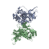
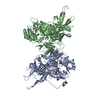
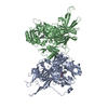
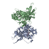
| Title | Crystal structure of box C/D RNP with 13 nt guide regions and 11 nt substrates | ||||||
 Components Components |
| ||||||
 Keywords Keywords | TRANSFERASE/RNA / 2'-O-methylation / guide RNA / RNP / TRANSFERASE-RNA complex | ||||||
| Function / homology |  Function and homology information Function and homology informationhistone H2AQ104 methyltransferase activity / box C/D sno(s)RNA 3'-end processing / rRNA methyltransferase activity / box C/D methylation guide snoRNP complex / rRNA methylation / ribonuclease P activity / tRNA 5'-leader removal / tRNA processing / snoRNA binding / Transferases; Transferring one-carbon groups; Methyltransferases ...histone H2AQ104 methyltransferase activity / box C/D sno(s)RNA 3'-end processing / rRNA methyltransferase activity / box C/D methylation guide snoRNP complex / rRNA methylation / ribonuclease P activity / tRNA 5'-leader removal / tRNA processing / snoRNA binding / Transferases; Transferring one-carbon groups; Methyltransferases / ribosome biogenesis / rRNA binding / structural constituent of ribosome / ribosome / translation / ribonucleoprotein complex / RNA binding / cytoplasm Similarity search - Function | ||||||
| Biological species |   Sulfolobus solfataricus (archaea) Sulfolobus solfataricus (archaea) | ||||||
| Method |  X-RAY DIFFRACTION / SYNCHROTRON / MOLECULAR REPLACEMENT / Resolution: 3.129 Å X-RAY DIFFRACTION / SYNCHROTRON / MOLECULAR REPLACEMENT / Resolution: 3.129 Å | ||||||
 Authors Authors | Yang, Z. / Lin, J. / Ye, K. | ||||||
 Citation Citation | Journal: Proc.Natl.Acad.Sci.USA / Year: 2016 Title: Box C/D guide RNAs recognize a maximum of 10 nt of substrates Authors: Yang, Z. / Lin, J. / Ye, K. | ||||||
| History |
|
- Structure visualization
| Structure viewer | Molecule: MolmilJmol/JSmol |
|---|
- Downloads & links
-Download
| PDBx/mmCIF format | 5gip.cif.gz | 659.6 KB | Display | PDBx/mmCIF format |
|---|---|---|---|---|
| PDB format | pdb5gip.ent.gz | 535.3 KB | Display | PDB format |
| PDBx/mmJSON format | 5gip.json.gz | Tree view | PDBx/mmJSON format | |
| Others |  Other downloads Other downloads |
-Validation report
| Arichive directory | https://data.pdbj.org/pub/pdb/validation_reports/gi/5gipftp://data.pdbj.org/pub/pdb/validation_reports/gi/5gip | HTTPS FTP |
|---|
-Related structure data
| Related structure data |  5ginC  5gioC  3plaS C: citing same article ( S: Starting model for refinement |
|---|---|
| Similar structure data |


-Links
 PDBj
PDBj


- Assembly
| Deposited unit | 
| ||||||||
|---|---|---|---|---|---|---|---|---|---|
| 1 | 
| ||||||||
| 2 | 
| ||||||||
| Unit cell |
|
-Components
-Protein , 3 types, 12 molecules ABKLCDMNEFOP
| #1: Protein | Mass: 44168.531 Da / Num. of mol.: 4 / Fragment: UNP residues 3-379 Source method: isolated from a genetically manipulated source Source: (gene. exp.) Sulfolobus solfataricus (archaea) / Gene: SULA_1947, SULB_1948, SULC_1946 / Production host:  #2: Protein | Mass: 14075.301 Da / Num. of mol.: 4 / Fragment: UNP residues 3-127 Source method: isolated from a genetically manipulated source Source: (gene. exp.) Sulfolobus solfataricus (archaea) / Gene: rpl7ae, SULA_1106, SULB_1107, SULC_1105 / Production host: #3: Protein | Mass: 26439.375 Da / Num. of mol.: 4 / Fragment: UNP residues 3-232 Source method: isolated from a genetically manipulated source Source: (gene. exp.) Sulfolobus solfataricus (archaea) / Gene: flpA, SSOP1_0970, SULA_1948, SULB_1949, SULC_1947 / Production host: References: UniProt: A0A0E3JUC9, UniProt: P58032*PLUS, Transferases; Transferring one-carbon groups; Methyltransferases |
|---|
-RNA chain , 2 types, 8 molecules GHQRIJST
| #4: RNA chain | Mass: 13197.862 Da / Num. of mol.: 4 / Source method: obtained synthetically / Source: (synth.) Sulfolobus solfataricus (archaea)#5: RNA chain | Mass: 3484.098 Da / Num. of mol.: 4 / Source method: obtained synthetically / Source: (synth.) Sulfolobus solfataricus (archaea) |
|---|
-Non-polymers , 1 types, 4 molecules 
| #6: Chemical | ChemComp-SAH /  Type: L-peptide linking / Mass: 384.411 Da / Num. of mol.: 4 / Source method: obtained synthetically / Formula: C14H20N6O5S Type: L-peptide linking / Mass: 384.411 Da / Num. of mol.: 4 / Source method: obtained synthetically / Formula: C14H20N6O5S |
|---|
-Experimental details
-Experiment
| Experiment | Method: X-RAY DIFFRACTION / Number of used crystals: 1 |
|---|
- Sample preparation
| Crystal | Density Matthews: 2.76 Å3/Da / Density % sol: 55.49 % |
|---|---|
| Crystal grow | Temperature: 293 K / Method: vapor diffusion, hanging drop Details: 1.7M DL-malic acid, 0.1M potassium sodium tartrate tetrahydrate |
-Data collection
| Diffraction | Mean temperature: 100 K |
|---|---|
| Diffraction source | Source: SYNCHROTRON / Site: SSRF  / Beamline: BL17U / Wavelength: 0.97915 Å / Beamline: BL17U / Wavelength: 0.97915 Å |
| Detector | Type: ADSC QUANTUM 315r / Detector: CCD / Date: May 11, 2013 |
| Radiation | Protocol: SINGLE WAVELENGTH / Monochromatic (M) / Laue (L): M / Scattering type: x-ray |
| Radiation wavelength | Wavelength: 0.97915 Å / Relative weight: 1 |
| Reflection | Resolution: 3.12→25 Å / Num. obs: 74336 / % possible obs: 97.3 % / Redundancy: 2.3 % / Rmerge(I) obs: 0.087 / Net I/σ(I): 22.5 |
| Reflection shell | Resolution: 3.13→3.21 Å |
- Processing
| Software |
| |||||||||||||||||||||||||||||||||||||||||||||||||||||||||||||||||||||||||||||||||||||||||||||||||||||||||
|---|---|---|---|---|---|---|---|---|---|---|---|---|---|---|---|---|---|---|---|---|---|---|---|---|---|---|---|---|---|---|---|---|---|---|---|---|---|---|---|---|---|---|---|---|---|---|---|---|---|---|---|---|---|---|---|---|---|---|---|---|---|---|---|---|---|---|---|---|---|---|---|---|---|---|---|---|---|---|---|---|---|---|---|---|---|---|---|---|---|---|---|---|---|---|---|---|---|---|---|---|---|---|---|---|---|---|
| Refinement | Method to determine structure: MOLECULAR REPLACEMENT Starting model: 3PLA Resolution: 3.129→24.859 Å / SU ML: 0.37 / Cross valid method: FREE R-VALUE / σ(F): 1.96 / Phase error: 25.72 / Stereochemistry target values: ML
| |||||||||||||||||||||||||||||||||||||||||||||||||||||||||||||||||||||||||||||||||||||||||||||||||||||||||
| Solvent computation | Shrinkage radii: 0.9 Å / VDW probe radii: 1.11 Å / Solvent model: FLAT BULK SOLVENT MODEL | |||||||||||||||||||||||||||||||||||||||||||||||||||||||||||||||||||||||||||||||||||||||||||||||||||||||||
| Refinement step | Cycle: LAST / Resolution: 3.129→24.859 Å
| |||||||||||||||||||||||||||||||||||||||||||||||||||||||||||||||||||||||||||||||||||||||||||||||||||||||||
| Refine LS restraints |
| |||||||||||||||||||||||||||||||||||||||||||||||||||||||||||||||||||||||||||||||||||||||||||||||||||||||||
| LS refinement shell |
|
