 Movie
Movie Controller


+ Open data

- Basic information
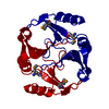
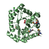
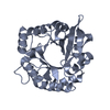
| Entry | Database: PDB / ID: 5fzr | ||||||
|---|---|---|---|---|---|---|---|
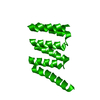
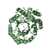
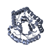
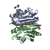
| Title | Designed TPR Protein M4N delta C (CF I) | ||||||
 Components Components | DESIGNED TPR PROTEIN | ||||||
 Keywords Keywords | UNKNOWN FUNCTION / TETRATRICOPEPTIDE REPEAT | ||||||
| Function / homology | Tetratricopeptide repeat domain / Serine Threonine Protein Phosphatase 5, Tetratricopeptide repeat / Alpha Horseshoe / Mainly Alpha Function and homology information Function and homology information | ||||||
| Biological species | SYNTHETIC CONSTRUCT (others) | ||||||
| Method |  X-RAY DIFFRACTION / SYNCHROTRON / MOLECULAR REPLACEMENT / Resolution: 2.045 Å X-RAY DIFFRACTION / SYNCHROTRON / MOLECULAR REPLACEMENT / Resolution: 2.045 Å | ||||||
 Authors Authors | Albrecht, R. / Zhu, H. / Hartmann, M.D. | ||||||
 Citation Citation | Journal: Elife / Year: 2016 Title: Origin of a folded repeat protein from an intrinsically disordered ancestor. Authors: Zhu, H. / Sepulveda, E. / Hartmann, M.D. / Kogenaru, M. / Ursinus, A. / Sulz, E. / Albrecht, R. / Coles, M. / Martin, J. / Lupas, A.N. | ||||||
| History |
|
- Structure visualization
| Structure viewer | Molecule: MolmilJmol/JSmol |
|---|
- Downloads & links
-Download
| PDBx/mmCIF format | 5fzr.cif.gz | 168.8 KB | Display | PDBx/mmCIF format |
|---|---|---|---|---|
| PDB format | pdb5fzr.ent.gz | 137.4 KB | Display | PDB format |
| PDBx/mmJSON format | 5fzr.json.gz | Tree view | PDBx/mmJSON format | |
| Others |  Other downloads Other downloads |
-Validation report
| Arichive directory | https://data.pdbj.org/pub/pdb/validation_reports/fz/5fzrftp://data.pdbj.org/pub/pdb/validation_reports/fz/5fzr | HTTPS FTP |
|---|
-Related structure data
| Related structure data |  5fzqSC  5fzsC S: Starting model for refinement C: citing same article ( |
|---|---|
| Similar structure data |

-Links
 PDBj
PDBj- Assembly
| Deposited unit | 
| ||||||||
|---|---|---|---|---|---|---|---|---|---|
| 1 | 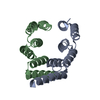
| ||||||||
| 2 | 
| ||||||||
| Unit cell |
|
-Components
| #1: Protein | Mass: 11831.779 Da / Num. of mol.: 4 Source method: isolated from a genetically manipulated source Source: (gene. exp.) SYNTHETIC CONSTRUCT (others) / Production host:  #2: Water | ChemComp-HOH / |  Mass: 18.015 Da / Num. of mol.: 150 / Source method: isolated from a natural source / Formula: H2O Mass: 18.015 Da / Num. of mol.: 150 / Source method: isolated from a natural source / Formula: H2O |
|---|
-Experimental details
-Experiment
| Experiment | Method: X-RAY DIFFRACTION / Number of used crystals: 1 |
|---|
- Sample preparation
| Crystal | Density Matthews: 2.1 Å3/Da / Density % sol: 42 % / Description: NONE |
|---|
-Data collection
| Diffraction | Mean temperature: 100 K |
|---|---|
| Diffraction source | Source: SYNCHROTRON / Site: SLS  / Beamline: X10SA / Wavelength: 1.071 / Beamline: X10SA / Wavelength: 1.071 |
| Detector | Type: DECTRIS PILATUS 6M / Detector: PIXEL / Date: Mar 22, 2014 |
| Radiation | Protocol: SINGLE WAVELENGTH / Monochromatic (M) / Laue (L): M / Scattering type: x-ray |
| Radiation wavelength | Wavelength: 1.071 Å / Relative weight: 1 |
| Reflection | Resolution: 2.05→38.3 Å / Num. obs: 25655 / % possible obs: 98.4 % / Observed criterion σ(I): -3 / Redundancy: 5.58 % / Biso Wilson estimate: 36.2 Å2 / Rmerge(I) obs: 0.06 / Net I/σ(I): 18.4 |
| Reflection shell | Resolution: 2.05→2.17 Å / Redundancy: 5.69 % / Rmerge(I) obs: 0.71 / Mean I/σ(I) obs: 2.1 / % possible all: 94.1 |
- Processing
| Software |
| |||||||||||||||||||||||||||||||||||||||||||||||||||||||||||||||||||||||||||||||||||||||||||||||||||||||||||||||||||||||||||||
|---|---|---|---|---|---|---|---|---|---|---|---|---|---|---|---|---|---|---|---|---|---|---|---|---|---|---|---|---|---|---|---|---|---|---|---|---|---|---|---|---|---|---|---|---|---|---|---|---|---|---|---|---|---|---|---|---|---|---|---|---|---|---|---|---|---|---|---|---|---|---|---|---|---|---|---|---|---|---|---|---|---|---|---|---|---|---|---|---|---|---|---|---|---|---|---|---|---|---|---|---|---|---|---|---|---|---|---|---|---|---|---|---|---|---|---|---|---|---|---|---|---|---|---|---|---|---|
| Refinement | Method to determine structure: MOLECULAR REPLACEMENT Starting model: PDB ENTRY 5FZQ Resolution: 2.045→38.252 Å / SU ML: 0.31 / σ(F): 1.36 / Phase error: 34.27 / Stereochemistry target values: ML
| |||||||||||||||||||||||||||||||||||||||||||||||||||||||||||||||||||||||||||||||||||||||||||||||||||||||||||||||||||||||||||||
| Solvent computation | Shrinkage radii: 0.9 Å / VDW probe radii: 1.11 Å / Solvent model: FLAT BULK SOLVENT MODEL | |||||||||||||||||||||||||||||||||||||||||||||||||||||||||||||||||||||||||||||||||||||||||||||||||||||||||||||||||||||||||||||
| Refinement step | Cycle: LAST / Resolution: 2.045→38.252 Å
| |||||||||||||||||||||||||||||||||||||||||||||||||||||||||||||||||||||||||||||||||||||||||||||||||||||||||||||||||||||||||||||
| Refine LS restraints |
| |||||||||||||||||||||||||||||||||||||||||||||||||||||||||||||||||||||||||||||||||||||||||||||||||||||||||||||||||||||||||||||
| LS refinement shell |
| |||||||||||||||||||||||||||||||||||||||||||||||||||||||||||||||||||||||||||||||||||||||||||||||||||||||||||||||||||||||||||||
| Refinement TLS params. | Method: refined / Refine-ID: X-RAY DIFFRACTION
| |||||||||||||||||||||||||||||||||||||||||||||||||||||||||||||||||||||||||||||||||||||||||||||||||||||||||||||||||||||||||||||
| Refinement TLS group |
|
