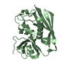
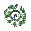
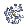
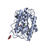
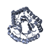
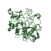
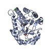
- PDB-2ook: Crystal structure of a protein with unknown function (YP_749275.1... -
+
Open data
ID or keywords:
Loading...
-
Basic information
Entry
Database: PDB / ID: 2ook
Title
Crystal structure of a protein with unknown function (YP_749275.1) from Shewanella Frigidimarina NCIMB 400 at 1.80 A resolution
Components
Hypothetical protein
Keywords
STRUCTURAL GENOMICS / UNKNOWN FUNCTION / YP_749275.1 / hypothetical protein / Joint Center for Structural Genomics / JCSG / Protein Structure Initiative / PSI-2
BIOMOLECULE: 1 THIS ENTRY CONTAINS THE CRYSTALLOGRAPHIC ASYMMETRIC UNIT WHICH CONSISTS OF 2 CHAINS. ... BIOMOLECULE: 1 THIS ENTRY CONTAINS THE CRYSTALLOGRAPHIC ASYMMETRIC UNIT WHICH CONSISTS OF 2 CHAINS. SEE REMARK 350 FOR INFORMATION ON GENERATING THE BIOLOGICAL MOLECULE(S). SIZE EXCLUSION CHROMATOGRAPHY WITH STATIC LIGHT SCATTERING SUPPORTS THE ASSIGNMENT OF A DIMER AS THE SIGNIFICANT OLIGOMERIZATION STATE.
Remark 999
SEQUENCE THE CONSTRUCT WAS EXPRESSED WITH A PURIFICATION TAG MGSDKIHHHHHHENLYFQG. THE TAG WAS ... SEQUENCE THE CONSTRUCT WAS EXPRESSED WITH A PURIFICATION TAG MGSDKIHHHHHHENLYFQG. THE TAG WAS REMOVED WITH TEV PROTEASE LEAVING ONLY A GLYCINE (0) FOLLOWED BY THE TARGET SEQUENCE.
Type: MARMOSAIC 325 mm CCD / Detector: CCD / Date: Jan 7, 2007 / Details: Flat mirror (vertical focusing)
Radiation
Monochromator: Single crystal Si(111) bent (horizontal focusing) Protocol: MAD / Monochromatic (M) / Laue (L): M / Scattering type: x-ray
Radiation wavelength
ID
Wavelength (Å)
Relative weight
1
0.91837
1
2
0.9791
1
3
0.97879
1
Reflection
Resolution: 1.8→28.571 Å / Num. obs: 22625 / % possible obs: 96.5 % / Redundancy: 3.8 % / Biso Wilson estimate: 24.85 Å2 / Rmerge(I) obs: 0.056 / Rsym value: 0.056 / Net I/σ(I): 13.6
Reflection shell
Diffraction-ID: 1
Resolution (Å)
Redundancy (%)
Rmerge(I) obs
Mean I/σ(I) obs
Num. measured all
Num. unique all
Rsym value
% possible all
1.8-1.85
3.8
0.653
2.1
6294
1640
0.653
95.3
1.85-1.9
3.8
0.514
1.5
6128
1597
0.514
95.5
1.9-1.95
3.8
0.407
1.9
5975
1570
0.407
96
1.95-2.01
3.8
0.294
2.6
5762
1507
0.294
95.7
2.01-2.08
3.8
0.226
3.3
5674
1487
0.226
96.3
2.08-2.15
3.8
0.184
4.2
5509
1436
0.184
96.2
2.15-2.23
3.8
0.146
5.3
5297
1379
0.146
96.2
2.23-2.32
3.8
0.126
5.9
5006
1315
0.126
96.5
2.32-2.43
3.8
0.105
7.1
4939
1292
0.105
96.8
2.43-2.55
3.8
0.092
8
4697
1232
0.092
96.6
2.55-2.68
3.8
0.075
9.2
4522
1188
0.075
96.9
2.68-2.85
3.8
0.063
11.3
4219
1104
0.063
97.1
2.85-3.04
3.8
0.051
13.6
4007
1054
0.051
97.5
3.04-3.29
3.8
0.038
17
3732
984
0.038
97.1
3.29-3.6
3.8
0.035
17.3
3375
891
0.035
97.5
3.6-4.02
3.8
0.031
20.4
3155
839
0.031
97.6
4.02-4.65
3.7
0.028
22.9
2686
722
0.028
97.9
4.65-5.69
3.7
0.03
19.8
2340
632
0.03
98
5.69-8.05
3.6
0.035
18.4
1771
492
0.035
98.4
8.05-28.57
3.4
0.029
23
908
264
0.029
93.5
-
Phasing
Phasing
Method: MAD
-
Processing
Software
Name
Version
Classification
NB
MolProbity
3beta29
modelbuilding
SHELX
phasing
REFMAC
5.2.0019
refinement
SCALA
datascaling
PDB_EXTRACT
2
dataextraction
MAR345
CCD
datacollection
MOSFLM
datareduction
CCP4
(SCALA)
datascaling
SHELXD
phasing
autoSHARP
phasing
Refinement
Method to determine structure: MAD / Resolution: 1.8→28.571 Å / Cor.coef. Fo:Fc: 0.964 / Cor.coef. Fo:Fc free: 0.939 / SU B: 6.712 / SU ML: 0.106 / TLS residual ADP flag: LIKELY RESIDUAL / Cross valid method: THROUGHOUT / σ(F): 0 / ESU R: 0.146 / ESU R Free: 0.141 Stereochemistry target values: MAXIMUM LIKELIHOOD WITH PHASES Details: 1. HYDROGENS HAVE BEEN ADDED IN THE RIDING POSITIONS. 2. A MET-INHIBITION PROTOCOL WAS USED FOR SELENOMETHIONINE INCORPORATION DURING PROTEIN EXPRESSION. THE OCCUPANCY OF THE SE ATOMS IN THE ...Details: 1. HYDROGENS HAVE BEEN ADDED IN THE RIDING POSITIONS. 2. A MET-INHIBITION PROTOCOL WAS USED FOR SELENOMETHIONINE INCORPORATION DURING PROTEIN EXPRESSION. THE OCCUPANCY OF THE SE ATOMS IN THE MSE RESIDUES WAS REDUCED TO 0.75 FOR THE REDUCED SCATTERING POWER DUE TO PARTIAL S-MET INCORPORATION. 3. RESIDUES 1 IN CHAIN A AND 1-2, 80-81 IN CHAIN B ARE DISORDERED AND NOT INCLUDED IN THE MODEL. 4. EDO MOLECULES FROM THE CRYO SOLUTION ARE MODELED. 5. ATOM RECORDS CONTAIN RESIDUAL B FACTORS ONLY.
Rfactor
Num. reflection
% reflection
Selection details
Rfree
0.233
1162
5.1 %
RANDOM
Rwork
0.183
-
-
-
all
0.186
-
-
-
obs
0.186
22625
96.02 %
-
Solvent computation
Ion probe radii: 0.8 Å / Shrinkage radii: 0.8 Å / VDW probe radii: 1.2 Å / Solvent model: BABINET MODEL WITH MASK
Displacement parameters
Biso mean: 29.171 Å2
Baniso -1
Baniso -2
Baniso -3
1-
-2.16 Å2
0 Å2
-1.2 Å2
2-
-
2.58 Å2
0 Å2
3-
-
-
-0.51 Å2
Refinement step
Cycle: LAST / Resolution: 1.8→28.571 Å
Protein
Nucleic acid
Ligand
Solvent
Total
Num. atoms
1963
0
24
224
2211
Refine LS restraints
Refine-ID
Type
Dev ideal
Dev ideal target
Number
X-RAY DIFFRACTION
r_bond_refined_d
0.012
0.022
2104
X-RAY DIFFRACTION
r_bond_other_d
0.001
0.02
1412
X-RAY DIFFRACTION
r_angle_refined_deg
1.526
1.953
2860
X-RAY DIFFRACTION
r_angle_other_deg
0.899
3
3460
X-RAY DIFFRACTION
r_dihedral_angle_1_deg
4.704
5
267
X-RAY DIFFRACTION
r_dihedral_angle_2_deg
32.917
24.946
93
X-RAY DIFFRACTION
r_dihedral_angle_3_deg
11.623
15
365
X-RAY DIFFRACTION
r_dihedral_angle_4_deg
13.645
15
6
X-RAY DIFFRACTION
r_chiral_restr
0.086
0.2
318
X-RAY DIFFRACTION
r_gen_planes_refined
0.005
0.02
2323
X-RAY DIFFRACTION
r_gen_planes_other
0.001
0.02
431
X-RAY DIFFRACTION
r_nbd_refined
0.209
0.2
381
X-RAY DIFFRACTION
r_nbd_other
0.199
0.2
1410
X-RAY DIFFRACTION
r_nbtor_refined
0.184
0.2
975
X-RAY DIFFRACTION
r_nbtor_other
0.086
0.2
1093
X-RAY DIFFRACTION
r_xyhbond_nbd_refined
0.178
0.2
192
X-RAY DIFFRACTION
r_symmetry_vdw_refined
0.249
0.2
14
X-RAY DIFFRACTION
r_symmetry_vdw_other
0.314
0.2
40
X-RAY DIFFRACTION
r_symmetry_hbond_refined
0.176
0.2
14
X-RAY DIFFRACTION
r_mcbond_it
2.073
3
1367
X-RAY DIFFRACTION
r_mcbond_other
0.524
3
518
X-RAY DIFFRACTION
r_mcangle_it
2.832
5
2020
X-RAY DIFFRACTION
r_scbond_it
4.445
8
972
X-RAY DIFFRACTION
r_scangle_it
5.68
11
828
LS refinement shell
Resolution: 1.8→1.847 Å / Total num. of bins used: 20
Rfactor
Num. reflection
% reflection
Rfree
0.306
74
-
Rwork
0.243
1567
-
obs
-
1641
94.91 %
Refinement TLS params.
Method: refined / Refine-ID: X-RAY DIFFRACTION
ID
L11 (°2)
L12 (°2)
L13 (°2)
L22 (°2)
L23 (°2)
L33 (°2)
S11 (Å °)
S12 (Å °)
S13 (Å °)
S21 (Å °)
S22 (Å °)
S23 (Å °)
S31 (Å °)
S32 (Å °)
S33 (Å °)
T11 (Å2)
T12 (Å2)
T13 (Å2)
T22 (Å2)
T23 (Å2)
T33 (Å2)
Origin x (Å)
Origin y (Å)
Origin z (Å)
1
3.3434
1.5389
-0.0728
1.9818
0.5879
3.3031
0.0797
-0.0643
0.066
0.0612
-0.0276
0.0962
-0.0599
-0.1041
-0.0522
-0.143
-0.0233
0.0113
-0.2224
0.015
-0.1556
14.942
20.651
17.926
2
4.3945
1.4246
0.7787
1.155
-0.2384
2.287
0.1099
-0.1311
0.1426
0.1397
-0.0566
-0.0272
-0.0914
0.021
-0.0533
-0.1119
-0.0423
0.0213
-0.1938
-0.0221
-0.1572
33.538
29.315
20.888
Refinement TLS group
Refine-ID: X-RAY DIFFRACTION / Selection: ALL
ID
Refine TLS-ID
Auth asym-ID
Label asym-ID
Auth seq-ID
Label seq-ID
1
1
A
A
2 - 126
3 - 127
2
2
B
B
3 - 79
4 - 80
3
2
B
B
82 - 126
83 - 127
+
About Yorodumi
-
News
-
Feb 9, 2022. New format data for meta-information of EMDB entries
New format data for meta-information of EMDB entries
Version 3 of the EMDB header file is now the official format.
The previous official version 1.9 will be removed from the archive.
In the structure databanks used in Yorodumi, some data are registered as the other names, "COVID-19 virus" and "2019-nCoV". Here are the details of the virus and the list of structure data.
Jan 31, 2019. EMDB accession codes are about to change! (news from PDBe EMDB page)
EMDB accession codes are about to change! (news from PDBe EMDB page)
The allocation of 4 digits for EMDB accession codes will soon come to an end. Whilst these codes will remain in use, new EMDB accession codes will include an additional digit and will expand incrementally as the available range of codes is exhausted. The current 4-digit format prefixed with “EMD-” (i.e. EMD-XXXX) will advance to a 5-digit format (i.e. EMD-XXXXX), and so on. It is currently estimated that the 4-digit codes will be depleted around Spring 2019, at which point the 5-digit format will come into force.
The EM Navigator/Yorodumi systems omit the EMD- prefix.
Related info.:Q: What is EMD? / ID/Accession-code notation in Yorodumi/EM Navigator
Yorodumi is a browser for structure data from EMDB, PDB, SASBDB, etc.
This page is also the successor to EM Navigator detail page, and also detail information page/front-end page for Omokage search.
The word "yorodu" (or yorozu) is an old Japanese word meaning "ten thousand". "mi" (miru) is to see.
Related info.:EMDB / PDB / SASBDB / Comparison of 3 databanks / Yorodumi Search / Aug 31, 2016. New EM Navigator & Yorodumi / Yorodumi Papers / Jmol/JSmol / Function and homology information / Changes in new EM Navigator and Yorodumi
 Movie
Movie Controller

 Yorodumi
 Open data
 Basic information
 Components
 Keywords
 Function and homology information
 Shewanella frigidimarina (bacteria)
 X-RAY DIFFRACTION /
  Authors
 Citation
 Structure visualization
 Downloads & links
 Other downloads

 PDBj
PDBj
 Assembly
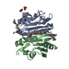

 Mass: 62.068 Da / Num. of mol.: 6 / Source method: obtained synthetically / Formula: C2H6O2
 Mass: 18.015 Da / Num. of mol.: 224 / Source method: isolated from a natural source / Formula: H2O
 Sample preparation
 / Beamline: BL11-1 / Wavelength: 0.91837, 0.97910, 0.97879
 Processing