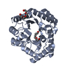
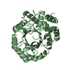
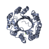
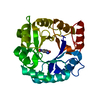
- PDB-2q02: Crystal structure of a xylose isomerase domain containing protein... -
+
Open data
ID or keywords:
Loading...
-
Basic information
Entry
Database: PDB / ID: 2q02
Title
Crystal structure of a xylose isomerase domain containing protein (stm4435) from salmonella typhimurium lt2 at 2.40 A resolution
Components
Putative cytoplasmic protein
Keywords
UNKNOWN FUNCTION / Putative cytoplasmic protein / structural genomics / Joint Center for Structural Genomics / JCSG / Protein Structure Initiative / PSI-2
SEQUENCE THE CONSTRUCT WAS EXPRESSED WITH A PURIFICATION TAG MGSDKIHHHHHHENLYFQG. THE TAG WAS ... SEQUENCE THE CONSTRUCT WAS EXPRESSED WITH A PURIFICATION TAG MGSDKIHHHHHHENLYFQG. THE TAG WAS REMOVED WITH TEV PROTEASE LEAVING ONLY A GLYCINE (0) FOLLOWED BY THE TARGET SEQUENCE.
Type: MARMOSAIC 325 mm CCD / Detector: CCD / Date: Feb 12, 2007 / Details: Flat mirror (vertical focusing)
Radiation
Monochromator: Single crystal Si(111) bent (horizontal focusing) Protocol: MAD / Monochromatic (M) / Laue (L): M / Scattering type: x-ray
Radiation wavelength
ID
Wavelength (Å)
Relative weight
1
0.91162
1
2
0.97922
1
3
0.97894
1
Reflection
Resolution: 2.4→29.386 Å / Num. obs: 48661 / % possible obs: 98.5 % / Observed criterion σ(I): -3 / Biso Wilson estimate: 44.32 Å2 / Rmerge(I) obs: 0.057 / Net I/σ(I): 9.83
Reflection shell
Diffraction-ID: 1
Resolution (Å)
Highest resolution (Å)
Rmerge(I) obs
Mean I/σ(I) obs
Num. measured obs
Num. unique obs
% possible all
2.4-2.49
0.322
2.4
15045
9080
92.4
2.49-2.58
0.283
2.8
14220
8428
99.8
2.58-2.7
0.237
3.6
15493
9522
98.8
2.7-2.84
0.188
4.3
15623
9202
99.8
2.84-3.02
0.137
5.8
16352
9539
99.8
3.02-3.25
0.102
7.8
15959
9264
99.7
3.25-3.58
0.069
11.2
15666
9398
99.1
3.58-4.09
0.048
15.9
14889
9176
98.3
4.09-5.14
0.033
20.8
16275
9354
99.7
5.14
0.025
23
16814
9424
98.4
-
Phasing
Phasing
Method: MAD
-
Processing
Software
Name
Version
Classification
NB
REFMAC
5.2.0005
refinement
PHENIX
refinement
SHELX
phasing
MolProbity
3beta29
modelbuilding
XSCALE
datascaling
PDB_EXTRACT
3
dataextraction
MAR345
CCD
datacollection
XDS
datareduction
SHELXD
phasing
autoSHARP
phasing
Refinement
Method to determine structure: MAD / Resolution: 2.4→29.386 Å / Cor.coef. Fo:Fc: 0.956 / Cor.coef. Fo:Fc free: 0.943 / SU B: 17.462 / SU ML: 0.206 / TLS residual ADP flag: LIKELY RESIDUAL / Cross valid method: THROUGHOUT / σ(F): 0 / ESU R: 0.407 / ESU R Free: 0.244 Stereochemistry target values: MAXIMUM LIKELIHOOD WITH PHASES Details: 1. HYDROGENS HAVE BEEN ADDED IN THE RIDING POSITIONS. 2. A MET-INHIBITION PROTOCOL WAS USED FOR SELENOMETHIONINE INCORPORATION DURING PROTEIN EXPRESSION. THE OCCUPANCY OF THE SE ATOMS IN THE ...Details: 1. HYDROGENS HAVE BEEN ADDED IN THE RIDING POSITIONS. 2. A MET-INHIBITION PROTOCOL WAS USED FOR SELENOMETHIONINE INCORPORATION DURING PROTEIN EXPRESSION. THE OCCUPANCY OF THE SE ATOMS IN THE MSE RESIDUES WAS REDUCED TO 0.75 FOR THE REDUCED SCATTERING POWER DUE TO PARTIAL S-MET INCORPORATION. 3. ATOM RECORD CONTAINS RESIDUAL B FACTORS ONLY. 4. DATA HAS PARTIAL-MEROHEDRAL TWINNING, WITH TWIN OPERATOR -K,-H,-L AND AN APPARENT TWIN FRACTION OF 10%. DATA WAS NOT DETWINNED DURING REFINEMENT. 5. THE R-FREE SET WAS GENERATED USING THE TWIN LAWS. 6. ZINC WAS MODELED BASED ON GEOMETRY AND COORDINATION ENVIRONMENT, AND CONFIRMED WITH X-RAY FLUORESCENCE AND ANOMALOUS DIFFERENCE FOURIER EXPERIMENTS. 7. CHLORINE ATOMS WERE MODELED BASED ON CRYSTALLIZATION CONDITIONS. 8. UNKNOWN LIGANDS (UNL) WERE MODELED BASED ON LOCATION OF PROPOSED ACTIVE SITE. 9. THERE IS AN UNMODELED DIFFERENCE DENSITY NEAR ARG A148.
Rfactor
Num. reflection
% reflection
Selection details
Rfree
0.228
2435
5 %
RANDOM
Rwork
0.184
-
-
-
obs
0.186
48605
99.3 %
-
Solvent computation
Ion probe radii: 0.8 Å / Shrinkage radii: 0.8 Å / VDW probe radii: 1.2 Å / Solvent model: MASK
Displacement parameters
Biso mean: 44.088 Å2
Baniso -1
Baniso -2
Baniso -3
1-
-1.36 Å2
0 Å2
0 Å2
2-
-
-1.38 Å2
0 Å2
3-
-
-
2.74 Å2
Refinement step
Cycle: LAST / Resolution: 2.4→29.386 Å
Protein
Nucleic acid
Ligand
Solvent
Total
Num. atoms
8552
0
27
236
8815
Refine LS restraints
Refine-ID
Type
Dev ideal
Dev ideal target
Number
X-RAY DIFFRACTION
r_bond_refined_d
0.014
0.022
8753
X-RAY DIFFRACTION
r_bond_other_d
0.002
0.02
8097
X-RAY DIFFRACTION
r_angle_refined_deg
1.333
1.968
11859
X-RAY DIFFRACTION
r_angle_other_deg
0.82
3
18722
X-RAY DIFFRACTION
r_dihedral_angle_1_deg
5.99
5
1095
X-RAY DIFFRACTION
r_dihedral_angle_2_deg
39.112
24.172
429
X-RAY DIFFRACTION
r_dihedral_angle_3_deg
14.895
15
1512
X-RAY DIFFRACTION
r_dihedral_angle_4_deg
20.184
15
73
X-RAY DIFFRACTION
r_chiral_restr
0.071
0.2
1354
X-RAY DIFFRACTION
r_gen_planes_refined
0.004
0.02
9820
X-RAY DIFFRACTION
r_gen_planes_other
0.001
0.02
1781
X-RAY DIFFRACTION
r_nbd_refined
0.214
0.2
1818
X-RAY DIFFRACTION
r_nbd_other
0.18
0.2
8319
X-RAY DIFFRACTION
r_nbtor_refined
0.175
0.2
4243
X-RAY DIFFRACTION
r_nbtor_other
0.084
0.2
5124
X-RAY DIFFRACTION
r_xyhbond_nbd_refined
0.151
0.2
245
X-RAY DIFFRACTION
r_metal_ion_refined
0.07
0.2
1
X-RAY DIFFRACTION
r_symmetry_vdw_refined
0.182
0.2
12
X-RAY DIFFRACTION
r_symmetry_vdw_other
0.228
0.2
65
X-RAY DIFFRACTION
r_symmetry_hbond_refined
0.104
0.2
9
X-RAY DIFFRACTION
r_mcbond_it
1.114
3
5975
X-RAY DIFFRACTION
r_mcbond_other
0.433
3
2207
X-RAY DIFFRACTION
r_mcangle_it
1.533
5
8766
X-RAY DIFFRACTION
r_scbond_it
3.504
8
3576
X-RAY DIFFRACTION
r_scangle_it
4.625
11
3087
Refine LS restraints NCS
Ens-ID: 1 / Refine-ID: X-RAY DIFFRACTION
Dom-ID
Auth asym-ID
Number
Type
Rms dev position (Å)
Weight position
1
A
948
TIGHTPOSITIONAL
0.05
0.05
2
B
948
TIGHTPOSITIONAL
0.04
0.05
3
C
948
TIGHTPOSITIONAL
0.05
0.05
4
D
948
TIGHTPOSITIONAL
0.05
0.05
1
A
2749
MEDIUMPOSITIONAL
0.28
0.5
2
B
2749
MEDIUMPOSITIONAL
0.3
0.5
3
C
2749
MEDIUMPOSITIONAL
0.26
0.5
4
D
2749
MEDIUMPOSITIONAL
0.3
0.5
1
A
948
TIGHTTHERMAL
0.1
0.5
2
B
948
TIGHTTHERMAL
0.11
0.5
3
C
948
TIGHTTHERMAL
0.11
0.5
4
D
948
TIGHTTHERMAL
0.1
0.5
1
A
2749
MEDIUMTHERMAL
0.63
2
2
B
2749
MEDIUMTHERMAL
0.71
2
3
C
2749
MEDIUMTHERMAL
0.64
2
4
D
2749
MEDIUMTHERMAL
0.65
2
LS refinement shell
Resolution: 2.4→2.461 Å / Total num. of bins used: 20
Rfactor
Num. reflection
% reflection
Rfree
0.308
186
-
Rwork
0.251
3230
-
obs
-
3416
96.44 %
Refinement TLS params.
Method: refined / Refine-ID: X-RAY DIFFRACTION
ID
L11 (°2)
L12 (°2)
L13 (°2)
L22 (°2)
L23 (°2)
L33 (°2)
S11 (Å °)
S12 (Å °)
S13 (Å °)
S21 (Å °)
S22 (Å °)
S23 (Å °)
S31 (Å °)
S32 (Å °)
S33 (Å °)
T11 (Å2)
T12 (Å2)
T13 (Å2)
T22 (Å2)
T23 (Å2)
T33 (Å2)
Origin x (Å)
Origin y (Å)
Origin z (Å)
1
2.6942
-0.5256
-0.3191
2.0677
-0.8819
1.5933
-0.022
-0.1685
0.0571
-0.035
-0.1191
-0.1228
0.2648
0.4098
0.1411
-0.0676
0.0921
0.0437
-0.0008
0.0589
-0.1185
67.062
84.187
36.06
2
1.5182
-0.2774
0.8421
3.5924
0.0163
2.0047
-0.1713
-0.0983
0.1349
-0.0023
0.0307
0.1307
-0.4175
-0.4919
0.1407
-0.0556
0.1095
-0.0386
-0.0398
-0.0369
-0.0966
58.709
90.734
67.114
3
1.6212
-0.0355
0.1024
1.9903
0.0449
1.2098
0.0029
-0.0957
-0.2691
0.2312
-0.0235
-0.0367
0.1783
0.085
0.0206
-0.1321
0.0178
0.0082
-0.1794
0.0168
-0.1008
52.445
46.545
65.169
4
2.922
0.4042
-0.3636
1.186
-0.1641
1.6362
-0.0772
0.3544
0.3497
-0.0855
0.0469
0.2071
-0.0495
-0.3304
0.0303
-0.1377
0.0154
-0.0084
-0.1001
0.0445
-0.0352
23.096
77.127
37.038
Refinement TLS group
ID
Refine-ID
Refine TLS-ID
Auth asym-ID
Label asym-ID
Auth seq-ID
Label seq-ID
1
X-RAY DIFFRACTION
1
A
A
10 - 271
11 - 272
2
X-RAY DIFFRACTION
2
B
B
0 - 271
1 - 272
3
X-RAY DIFFRACTION
3
C
C
1 - 271
2 - 272
4
X-RAY DIFFRACTION
4
D
D
0 - 271
1 - 272
+
About Yorodumi
-
News
-
Feb 9, 2022. New format data for meta-information of EMDB entries
New format data for meta-information of EMDB entries
Version 3 of the EMDB header file is now the official format.
The previous official version 1.9 will be removed from the archive.
In the structure databanks used in Yorodumi, some data are registered as the other names, "COVID-19 virus" and "2019-nCoV". Here are the details of the virus and the list of structure data.
Jan 31, 2019. EMDB accession codes are about to change! (news from PDBe EMDB page)
EMDB accession codes are about to change! (news from PDBe EMDB page)
The allocation of 4 digits for EMDB accession codes will soon come to an end. Whilst these codes will remain in use, new EMDB accession codes will include an additional digit and will expand incrementally as the available range of codes is exhausted. The current 4-digit format prefixed with “EMD-” (i.e. EMD-XXXX) will advance to a 5-digit format (i.e. EMD-XXXXX), and so on. It is currently estimated that the 4-digit codes will be depleted around Spring 2019, at which point the 5-digit format will come into force.
The EM Navigator/Yorodumi systems omit the EMD- prefix.
Related info.:Q: What is EMD? / ID/Accession-code notation in Yorodumi/EM Navigator
Yorodumi is a browser for structure data from EMDB, PDB, SASBDB, etc.
This page is also the successor to EM Navigator detail page, and also detail information page/front-end page for Omokage search.
The word "yorodu" (or yorozu) is an old Japanese word meaning "ten thousand". "mi" (miru) is to see.
Related info.:EMDB / PDB / SASBDB / Comparison of 3 databanks / Yorodumi Search / Aug 31, 2016. New EM Navigator & Yorodumi / Yorodumi Papers / Jmol/JSmol / Function and homology information / Changes in new EM Navigator and Yorodumi
 Movie
Movie Controller

 Yorodumi
 Open data
 Basic information
 Components
 Keywords
 Function and homology information
 Salmonella typhimurium LT2 (bacteria)
 X-RAY DIFFRACTION /
  Authors
 Citation
 Structure visualization
 Downloads & links
 Other downloads
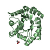


 PDBj
PDBj


 Assembly

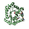


 Mass: 65.409 Da / Num. of mol.: 6 / Source method: obtained synthetically / Formula: Zn
 Mass: 35.453 Da / Num. of mol.: 8 / Source method: obtained synthetically / Formula: Cl
 Mass: 18.015 Da / Num. of mol.: 236 / Source method: isolated from a natural source / Formula: H2O
 Sample preparation
 / Beamline: BL11-1 / Wavelength: 0.91162, 0.97922, 0.97894
 Processing