 Movie
Movie Controller

[English] 日本語
 Yorodumi
- PDB-5fn3: Cryo-EM structure of gamma secretase in class 1 of the apo- state... -
+ Open data

- Basic information
| Entry | Database: PDB / ID: 5fn3 | ||||||
|---|---|---|---|---|---|---|---|
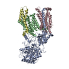
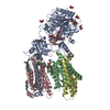
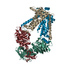
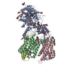
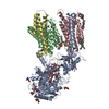
| Title | Cryo-EM structure of gamma secretase in class 1 of the apo- state ensemble | ||||||
 Components Components |
| ||||||
 Keywords Keywords | HYDROLASE | ||||||
| Function / homology |  Function and homology information Function and homology informationCajal-Retzius cell differentiation / positive regulation of L-glutamate import across plasma membrane / amyloid precursor protein biosynthetic process / positive regulation of endopeptidase activity / gamma-secretase complex / aspartic endopeptidase activity, intramembrane cleaving / positive regulation of amyloid precursor protein biosynthetic process / smooth endoplasmic reticulum calcium ion homeostasis / Noncanonical activation of NOTCH3 / protein catabolic process at postsynapse ...Cajal-Retzius cell differentiation / positive regulation of L-glutamate import across plasma membrane / amyloid precursor protein biosynthetic process / positive regulation of endopeptidase activity / gamma-secretase complex / aspartic endopeptidase activity, intramembrane cleaving / positive regulation of amyloid precursor protein biosynthetic process / smooth endoplasmic reticulum calcium ion homeostasis / Noncanonical activation of NOTCH3 / protein catabolic process at postsynapse / TGFBR3 PTM regulation / : / : / Notch receptor processing / positive regulation of coagulation / negative regulation of axonogenesis / membrane protein intracellular domain proteolysis / short-term synaptic potentiation / skin morphogenesis / T cell activation involved in immune response / choline transport / central nervous system myelination / NOTCH4 Activation and Transmission of Signal to the Nucleus / ciliary rootlet / regulation of resting membrane potential / neural retina development / Regulated proteolysis of p75NTR / myeloid dendritic cell differentiation / metanephros development / L-glutamate import across plasma membrane / endoplasmic reticulum calcium ion homeostasis / brain morphogenesis / amyloid precursor protein metabolic process / regulation of long-term synaptic potentiation / locomotion / cell fate specification / astrocyte activation involved in immune response / regulation of synaptic vesicle cycle / embryonic limb morphogenesis / regulation of postsynapse organization / myeloid cell homeostasis / aggresome / regulation of canonical Wnt signaling pathway / G protein-coupled dopamine receptor signaling pathway / skeletal system morphogenesis / Hydrolases; Acting on peptide bonds (peptidases); Aspartic endopeptidases / growth factor receptor binding / Golgi cisterna membrane / azurophil granule membrane / positive regulation of amyloid fibril formation / dorsal/ventral neural tube patterning / glutamate receptor signaling pathway / amyloid-beta formation / amyloid precursor protein catabolic process / mitochondrial transport / blood vessel development / regulation of neuron projection development / heart looping / positive regulation of dendritic spine development / cerebral cortex cell migration / smooth endoplasmic reticulum / membrane protein ectodomain proteolysis / adult behavior / positive regulation of receptor recycling / nuclear outer membrane / negative regulation of epidermal growth factor receptor signaling pathway / negative regulation of apoptotic signaling pathway / EPH-ephrin mediated repulsion of cells / T cell proliferation / hematopoietic progenitor cell differentiation / somitogenesis / Nuclear signaling by ERBB4 / negative regulation of ubiquitin-dependent protein catabolic process / neuron development / endopeptidase activator activity / autophagosome assembly / regulation of synaptic transmission, glutamatergic / calcium ion homeostasis / Degradation of the extracellular matrix / Notch signaling pathway / neuron projection maintenance / rough endoplasmic reticulum / epithelial cell proliferation / astrocyte activation / cellular response to calcium ion / NOTCH2 Activation and Transmission of Signal to the Nucleus / cerebellum development / thymus development / positive regulation of glycolytic process / NRIF signals cell death from the nucleus / Activated NOTCH1 Transmits Signal to the Nucleus / dendritic shaft / post-embryonic development / PDZ domain binding / neuromuscular junction / NOTCH3 Activation and Transmission of Signal to the Nucleus / apoptotic signaling pathway / neuron cellular homeostasis / protein processing / cell-cell adhesion Similarity search - Function | ||||||
| Biological species |  Homo sapiens (human)HOMO SAPIENS (human) Homo sapiens (human)HOMO SAPIENS (human) | ||||||
| Method | ELECTRON MICROSCOPY / single particle reconstruction / cryo EM / Resolution: 4.1 Å | ||||||
 Authors Authors | Bai, X.C. / Rajendra, E. / Yang, G.H. / Shi, Y.G. / Scheres, S.H.W. | ||||||
 Citation Citation | Journal: Elife / Year: 2015 Title: Sampling the conformational space of the catalytic subunit of human γ-secretase. Authors: Xiao-chen Bai / Eeson Rajendra / Guanghui Yang / Yigong Shi / Sjors H W Scheres /   Abstract: Human γ-secretase is an intra-membrane protease that cleaves many different substrates. Aberrant cleavage of Notch is implicated in cancer, while abnormalities in cutting amyloid precursor protein ...Human γ-secretase is an intra-membrane protease that cleaves many different substrates. Aberrant cleavage of Notch is implicated in cancer, while abnormalities in cutting amyloid precursor protein lead to Alzheimer's disease. Our previous cryo-EM structure of γ-secretase revealed considerable disorder in its catalytic subunit presenilin. Here, we describe an image classification procedure that characterizes molecular plasticity at the secondary structure level, and apply this method to identify three distinct conformations in our previous sample. In one of these conformations, an additional transmembrane helix is visible that cannot be attributed to the known components of γ-secretase. In addition, we present a γ-secretase structure in complex with the dipeptidic inhibitor N-[N-(3,5-difluorophenacetyl)-L-alanyl]-S-phenylglycine t-butyl ester (DAPT). Our results reveal how conformational mobility in the second and sixth transmembrane helices of presenilin is greatly reduced upon binding of DAPT or the additional helix, and form the basis for a new model of how substrate enters the transmembrane domain. | ||||||
| History |
|
- Structure visualization
| Movie |
Movie viewer |
|---|---|
| Structure viewer | Molecule: MolmilJmol/JSmol |
- Downloads & links
-Download
| PDBx/mmCIF format | 5fn3.cif.gz | 259 KB | Display | PDBx/mmCIF format |
|---|---|---|---|---|
| PDB format | pdb5fn3.ent.gz | 202 KB | Display | PDB format |
| PDBx/mmJSON format | 5fn3.json.gz | Tree view | PDBx/mmJSON format | |
| Others |  Other downloads Other downloads |
-Validation report
| Arichive directory | https://data.pdbj.org/pub/pdb/validation_reports/fn/5fn3ftp://data.pdbj.org/pub/pdb/validation_reports/fn/5fn3 | HTTPS FTP |
|---|
-Related structure data
| Related structure data | 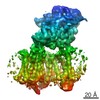 3238MC  3237C 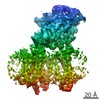 3239C 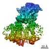 3240C 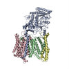 5fn2C 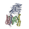 5fn4C 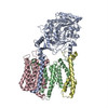 5fn5C C: citing same article ( M: map data used to model this data |
|---|---|
| Similar structure data |


-Links
 PDBj
PDBj


- Assembly
| Deposited unit | 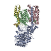
|
|---|---|
| 1 |
|
-Components
| #1: Protein | Mass: 78483.570 Da / Num. of mol.: 1 Source method: isolated from a genetically manipulated source Source: (gene. exp.) Homo sapiens (human) / Cell line: HEK293F / Gene: NCSTN, KIAA0253, UNQ1874/PRO4317 / Plasmid: PMLINK / Production host: HOMO SAPIENS (human) / References: UniProt: Q92542 |
|---|---|
| #2: Protein | Mass: 52651.465 Da / Num. of mol.: 1 Source method: isolated from a genetically manipulated source Source: (gene. exp.) Homo sapiens (human) / Cell line: HEK293F / Gene: PSEN1, AD3, PS1, PSNL1 / Plasmid: PMLINK / Production host: HOMO SAPIENS (human)References: UniProt: P49768, Hydrolases; Acting on peptide bonds (peptidases); Aspartic endopeptidases |
| #3: Protein | Mass: 29017.943 Da / Num. of mol.: 1 Source method: isolated from a genetically manipulated source Source: (gene. exp.) Homo sapiens (human) / Cell line: HEK293F / Gene: APH1A, PSF, CGI-78, UNQ579/PRO1141 / Plasmid: PMLINK / Production host: HOMO SAPIENS (human) / References: UniProt: Q96BI3 |
| #4: Protein | Mass: 12038.029 Da / Num. of mol.: 1 Source method: isolated from a genetically manipulated source Source: (gene. exp.) Homo sapiens (human) / Cell line: HEK293F / Gene: PSENEN, PEN2, MDS033 / Plasmid: PMLINK / Production host: HOMO SAPIENS (human) / References: UniProt: Q9NZ42 |
| #5: Protein/peptide | Mass: 1723.883 Da / Num. of mol.: 1 Source method: isolated from a genetically manipulated source Source: (gene. exp.) HOMO SAPIENS (human) / Cell line: HEK293F / Plasmid: PMLINK / Production host: HOMO SAPIENS (human) |
| Has protein modification | Y |
-Experimental details
-Experiment
| Experiment | Method: ELECTRON MICROSCOPY |
|---|---|
| EM experiment | Aggregation state: PARTICLE / 3D reconstruction method: single particle reconstruction |
- Sample preparation
| Component | Name: GAMMA SECRETASE / Type: COMPLEX |
|---|---|
| Buffer solution | Name: 25 MM HEPES, PH 7.4, 150 MM NACL AND AMPHIPOL A8-35 / pH: 7.4 Details: 25 MM HEPES, PH 7.4, 150 MM NACL AND AMPHIPOL A8-35 |
| Specimen | Conc.: 6 mg/ml / Embedding applied: NO / Shadowing applied: NO / Staining applied: NO / Vitrification applied: YES |
| Specimen support | Details: HOLEY CARBON |
| Vitrification | Instrument: FEI VITROBOT MARK IV / Cryogen name: ETHANE / Details: LIQUID ETHANE |
- Electron microscopy imaging
| Experimental equipment |  Model: Titan Krios / Image courtesy: FEI Company |
|---|---|
| Microscopy | Model: FEI TITAN KRIOS / Date: Oct 25, 2014 |
| Electron gun | Electron source:  FIELD EMISSION GUN / Accelerating voltage: 300 kV / Illumination mode: FLOOD BEAM FIELD EMISSION GUN / Accelerating voltage: 300 kV / Illumination mode: FLOOD BEAM |
| Electron lens | Mode: BRIGHT FIELD / Nominal magnification: 81000 X / Calibrated magnification: 35714 X / Nominal defocus max: 3200 nm / Nominal defocus min: 700 nm / Cs: 2.7 mm |
| Specimen holder | Temperature: 85 K |
| Image recording | Electron dose: 38 e/Å2 / Film or detector model: GATAN K2 QUANTUM (4k x 4k) |
| Image scans | Num. digital images: 2000 |
- Processing
| Symmetry | Point symmetry: C1 (asymmetric) | ||||||||||||
|---|---|---|---|---|---|---|---|---|---|---|---|---|---|
| 3D reconstruction | Resolution: 4.1 Å / Resolution method: FSC 0.143 CUT-OFF / Num. of particles: 63873 / Refinement type: HALF-MAPS REFINED INDEPENDENTLY / Symmetry type: POINT | ||||||||||||
| Refinement | Highest resolution: 4.1 Å | ||||||||||||
| Refinement step | Cycle: LAST / Highest resolution: 4.1 Å
|
