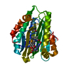
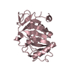
TRANSPORT PROTEIN / INTRAFLAGELLAR TRANSPORT / IFTB / IFT52 / PROTEIN-PROTEIN INTERACTION
Function / homology
Function and homology information
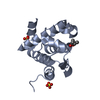
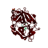
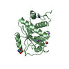
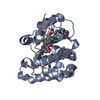
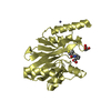
photoreceptor cell cilium / dendrite terminus / intraciliary anterograde transport / intraciliary transport particle B / Intraflagellar transport / neural tube formation / intraciliary transport / photoreceptor connecting cilium / Hedgehog 'off' state / dorsal/ventral pattern formation ...photoreceptor cell cilium / dendrite terminus / intraciliary anterograde transport / intraciliary transport particle B / Intraflagellar transport / neural tube formation / intraciliary transport / photoreceptor connecting cilium / Hedgehog 'off' state / dorsal/ventral pattern formation / non-motile cilium assembly / determination of left/right symmetry / regulation of protein processing / embryonic digit morphogenesis / smoothened signaling pathway / heart looping / ciliary base / keratinocyte proliferation / cilium assembly / negative regulation of keratinocyte proliferation / ciliary tip / centriole / cilium / ciliary basal body / centrosome Similarity search - Function
ABC-type uncharacterised transport system / ABC-type uncharacterized transport system / Intraflagellar transport protein 52 homolog / : / Intraflagellar transport protein 52, C-terminal domain Similarity search - Domain/homology
In the structure databanks used in Yorodumi, some data are registered as the other names, "COVID-19 virus" and "2019-nCoV". Here are the details of the virus and the list of structure data.
Jan 31, 2019. EMDB accession codes are about to change! (news from PDBe EMDB page)
EMDB accession codes are about to change! (news from PDBe EMDB page)
The allocation of 4 digits for EMDB accession codes will soon come to an end. Whilst these codes will remain in use, new EMDB accession codes will include an additional digit and will expand incrementally as the available range of codes is exhausted. The current 4-digit format prefixed with “EMD-” (i.e. EMD-XXXX) will advance to a 5-digit format (i.e. EMD-XXXXX), and so on. It is currently estimated that the 4-digit codes will be depleted around Spring 2019, at which point the 5-digit format will come into force.
The EM Navigator/Yorodumi systems omit the EMD- prefix.
Related info.:Q: What is EMD? / ID/Accession-code notation in Yorodumi/EM Navigator
Yorodumi is a browser for structure data from EMDB, PDB, SASBDB, etc.
This page is also the successor to EM Navigator detail page, and also detail information page/front-end page for Omokage search.
The word "yorodu" (or yorozu) is an old Japanese word meaning "ten thousand". "mi" (miru) is to see.
Related info.:EMDB / PDB / SASBDB / Comparison of 3 databanks / Yorodumi Search / Aug 31, 2016. New EM Navigator & Yorodumi / Yorodumi Papers / Jmol/JSmol / Function and homology information / Changes in new EM Navigator and Yorodumi
 Movie
Movie Controller


 Open data
 Basic information
 Components
 Keywords
 Function and homology information
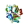
 X-RAY DIFFRACTION /
  Authors
 Citation
 Structure visualization
 Downloads & links
 Other downloads


 PDBj
PDBj

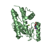
 Assembly

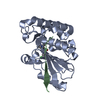
 Sample preparation
 / Beamline: X06DA / Wavelength: 1
  Processing