Monochromator: SI(111) MONOCHROMATOR / Protocol: SINGLE WAVELENGTH / Monochromatic (M) / Laue (L): M / Scattering type: x-ray
Radiation wavelength
Wavelength: 0.978946 Å / Relative weight: 1
Reflection
Resolution: 1.55→40.18 Å / Num. obs: 33089 / % possible obs: 99.6 % / Observed criterion σ(I): 0 / Redundancy: 3.7 % / Biso Wilson estimate: 16.03 Å2 / Rmerge(I) obs: 0.06 / Net I/σ(I): 11.53
Reflection shell
Resolution: 1.55→1.59 Å / Redundancy: 3.2 % / Rmerge(I) obs: 0.34 / Mean I/σ(I) obs: 2.91 / % possible all: 99.5
-
Processing
Software
Name
Version
Classification
PHENIX
(PHENIX.REFINE)
refinement
XDS
datareduction
XSCALE
datascaling
SHARP
phasing
Refinement
Method to determine structure: SAD Starting model: NONE Resolution: 1.55→40.185 Å / SU ML: 0.17 / σ(F): 2 / Phase error: 21.61 / Stereochemistry target values: ML Details: RESIDUES 128 AND 129 WERE NOT MODELLED DUE TO MISSING DENSITY
Rfactor
Num. reflection
% reflection
Rfree
0.2247
1655
5 %
Rwork
0.1791
-
-
obs
0.1813
33086
99.68 %
Solvent computation
Shrinkage radii: 0.9 Å / VDW probe radii: 1.11 Å / Solvent model: FLAT BULK SOLVENT MODEL / Bsol: 40.321 Å2 / ksol: 0.343 e/Å3
Displacement parameters
Biso mean: 21.47 Å2
Baniso -1
Baniso -2
Baniso -3
1-
2.9508 Å2
0 Å2
1.6075 Å2
2-
-
2.1651 Å2
-0 Å2
3-
-
-
-5.1159 Å2
Refinement step
Cycle: LAST / Resolution: 1.55→40.185 Å
Protein
Nucleic acid
Ligand
Solvent
Total
Num. atoms
1914
0
43
322
2279
Refine LS restraints
Refine-ID
Type
Dev ideal
Number
X-RAY DIFFRACTION
f_bond_d
0.007
2038
X-RAY DIFFRACTION
f_angle_d
1.164
2773
X-RAY DIFFRACTION
f_dihedral_angle_d
16.079
728
X-RAY DIFFRACTION
f_chiral_restr
0.083
272
X-RAY DIFFRACTION
f_plane_restr
0.006
362
LS refinement shell
Resolution (Å)
Rfactor Rfree
Num. reflection Rfree
Rfactor Rwork
Num. reflection Rwork
Refine-ID
% reflection obs (%)
1.5499-1.5955
0.2554
137
0.2193
2601
X-RAY DIFFRACTION
99
1.5955-1.647
0.2935
138
0.2141
2620
X-RAY DIFFRACTION
100
1.647-1.7059
0.2448
136
0.2041
2592
X-RAY DIFFRACTION
100
1.7059-1.7742
0.2377
137
0.192
2587
X-RAY DIFFRACTION
100
1.7742-1.8549
0.237
137
0.1836
2608
X-RAY DIFFRACTION
100
1.8549-1.9527
0.2427
137
0.189
2610
X-RAY DIFFRACTION
100
1.9527-2.075
0.2297
139
0.18
2630
X-RAY DIFFRACTION
100
2.075-2.2352
0.237
137
0.1786
2618
X-RAY DIFFRACTION
100
2.2352-2.4601
0.2139
137
0.1757
2599
X-RAY DIFFRACTION
100
2.4601-2.8161
0.2419
138
0.1837
2616
X-RAY DIFFRACTION
99
2.8161-3.5476
0.2155
139
0.1671
2647
X-RAY DIFFRACTION
99
3.5476-40.1986
0.1902
143
0.1573
2703
X-RAY DIFFRACTION
100
+
About Yorodumi
-
News
-
Feb 9, 2022. New format data for meta-information of EMDB entries
New format data for meta-information of EMDB entries
Version 3 of the EMDB header file is now the official format.
The previous official version 1.9 will be removed from the archive.
In the structure databanks used in Yorodumi, some data are registered as the other names, "COVID-19 virus" and "2019-nCoV". Here are the details of the virus and the list of structure data.
Jan 31, 2019. EMDB accession codes are about to change! (news from PDBe EMDB page)
EMDB accession codes are about to change! (news from PDBe EMDB page)
The allocation of 4 digits for EMDB accession codes will soon come to an end. Whilst these codes will remain in use, new EMDB accession codes will include an additional digit and will expand incrementally as the available range of codes is exhausted. The current 4-digit format prefixed with “EMD-” (i.e. EMD-XXXX) will advance to a 5-digit format (i.e. EMD-XXXXX), and so on. It is currently estimated that the 4-digit codes will be depleted around Spring 2019, at which point the 5-digit format will come into force.
The EM Navigator/Yorodumi systems omit the EMD- prefix.
Related info.:Q: What is EMD? / ID/Accession-code notation in Yorodumi/EM Navigator
Yorodumi is a browser for structure data from EMDB, PDB, SASBDB, etc.
This page is also the successor to EM Navigator detail page, and also detail information page/front-end page for Omokage search.
The word "yorodu" (or yorozu) is an old Japanese word meaning "ten thousand". "mi" (miru) is to see.
Related info.:EMDB / PDB / SASBDB / Comparison of 3 databanks / Yorodumi Search / Aug 31, 2016. New EM Navigator & Yorodumi / Yorodumi Papers / Jmol/JSmol / Function and homology information / Changes in new EM Navigator and Yorodumi
 Movie
Movie Controller

 Yorodumi
 Open data
 Basic information
 Components
 Keywords
 Function and homology information
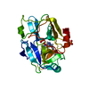
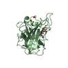
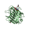
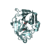
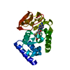
 SYNECHOCOCCUS SP. (bacteria)
 X-RAY DIFFRACTION /
  Authors
 Citation
 Structure visualization
 Downloads & links
 Other downloads
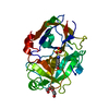


 PDBj
PDBj Assembly
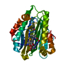

 Mass: 582.646 Da / Num. of mol.: 1 / Source method: obtained synthetically / Formula: C33H34N4O6
 Mass: 18.015 Da / Num. of mol.: 322 / Source method: isolated from a natural source / Formula: H2O
 Sample preparation
 / Beamline: X10SA / Wavelength: 0.978946
  Processing