 Movie
Movie Controller


+ Open data

- Basic information
| Entry | Database: PDB / ID: 5f0j | ||||||||||||
|---|---|---|---|---|---|---|---|---|---|---|---|---|---|
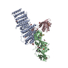
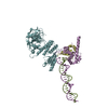
| Title | Structure of retromer VPS26-VPS35 subunits bound to SNX3 | ||||||||||||
 Components Components |
| ||||||||||||
 Keywords Keywords | TRANSPORT PROTEIN / protein transport / retromer / sorting nexin | ||||||||||||
| Function / homology |  Function and homology information Function and homology informationnegative regulation of early endosome to late endosome transport / negative regulation of protein transport / late endosome to Golgi transport / neurotransmitter receptor transport, endosome to plasma membrane / protein to membrane docking / negative regulation of protein localization / membrane invagination / mitochondrion-derived vesicle / negative regulation of protein homooligomerization / tubular endosome ...negative regulation of early endosome to late endosome transport / negative regulation of protein transport / late endosome to Golgi transport / neurotransmitter receptor transport, endosome to plasma membrane / protein to membrane docking / negative regulation of protein localization / membrane invagination / mitochondrion-derived vesicle / negative regulation of protein homooligomerization / tubular endosome / regulation of terminal button organization / regulation of dendritic spine maintenance / positive regulation of Wnt protein secretion / retromer, cargo-selective complex / mitochondrion to lysosome vesicle-mediated transport / intralumenal vesicle formation / WNT ligand biogenesis and trafficking / negative regulation of lysosomal protein catabolic process / negative regulation of late endosome to lysosome transport / retromer complex binding / positive regulation of dopamine receptor signaling pathway / positive regulation of locomotion involved in locomotory behavior / vesicle-mediated transport in synapse / neurotransmitter receptor transport, endosome to postsynaptic membrane / positive regulation of dopamine biosynthetic process / phosphatidylinositol-5-phosphate binding / protein localization to endosome / retromer complex / mitochondrial fragmentation involved in apoptotic process / voluntary musculoskeletal movement / transcytosis / host-mediated suppression of symbiont invasion / regulation of protein metabolic process / dopaminergic synapse / phosphatidylinositol-3-phosphate binding / endocytic recycling / early phagosome / regulation of synapse maturation / regulation of mitochondrion organization / phosphatidylinositol-4-phosphate binding / regulation of Wnt signaling pathway / phosphatidylinositol-3,5-bisphosphate binding / retrograde transport, endosome to Golgi / clathrin-coated vesicle / positive regulation of protein localization to cell periphery / positive regulation of mitochondrial fission / lysosome organization / negative regulation of phagocytosis / regulation of postsynapse assembly / regulation of macroautophagy / D1 dopamine receptor binding / regulation of presynapse assembly / response to bacterium / regulation of protein stability / positive regulation of neuron projection development / negative regulation of protein catabolic process / intracellular protein transport / protein destabilization / modulation of chemical synaptic transmission / negative regulation of inflammatory response / Wnt signaling pathway / positive regulation of protein catabolic process / late endosome / positive regulation of canonical Wnt signaling pathway / protein transport / presynapse / early endosome membrane / protein phosphatase binding / vesicle / early endosome / lysosome / endosome / endosome membrane / neuron projection / postsynaptic density / Ub-specific processing proteases / negative regulation of gene expression / lysosomal membrane / neuronal cell body / positive regulation of gene expression / perinuclear region of cytoplasm / glutamatergic synapse / extracellular exosome / cytoplasm / cytosol Similarity search - Function | ||||||||||||
| Biological species |  Homo sapiens (human) Homo sapiens (human) | ||||||||||||
| Method |  X-RAY DIFFRACTION / SYNCHROTRON / MOLECULAR REPLACEMENT / Resolution: 2.7 Å X-RAY DIFFRACTION / SYNCHROTRON / MOLECULAR REPLACEMENT / Resolution: 2.7 Å | ||||||||||||
 Authors Authors | Lucas, M. / Gershlick, D. / Vidaurrazaga, A. / Rojas, A.L. / Bonifacino, J.S. / Hierro, A. | ||||||||||||
| Funding support |  Spain, 3items Spain, 3items
| ||||||||||||
 Citation Citation | Journal: Cell / Year: 2016 Title: Structural Mechanism for Cargo Recognition by the Retromer Complex. Authors: Lucas, M. / Gershlick, D.C. / Vidaurrazaga, A. / Rojas, A.L. / Bonifacino, J.S. / Hierro, A. | ||||||||||||
| History |
|
- Structure visualization
| Structure viewer | Molecule: MolmilJmol/JSmol |
|---|
- Downloads & links
-Download
| PDBx/mmCIF format | 5f0j.cif.gz | 209.5 KB | Display | PDBx/mmCIF format |
|---|---|---|---|---|
| PDB format | pdb5f0j.ent.gz | 162.1 KB | Display | PDB format |
| PDBx/mmJSON format | 5f0j.json.gz | Tree view | PDBx/mmJSON format | |
| Others |  Other downloads Other downloads |
-Validation report
| Arichive directory | https://data.pdbj.org/pub/pdb/validation_reports/f0/5f0jftp://data.pdbj.org/pub/pdb/validation_reports/f0/5f0j | HTTPS FTP |
|---|
-Related structure data
| Related structure data |  5f0kC  5f0lC  5f0mC  5f0pC  2fauS  2ypsS S: Starting model for refinement C: citing same article ( |
|---|---|
| Similar structure data |


-Links
 PDBj
PDBj- Assembly
| Deposited unit | 
| ||||||||
|---|---|---|---|---|---|---|---|---|---|
| 1 |
| ||||||||
| Unit cell |
|
-Components
-Vacuolar protein sorting-associated protein ... , 2 types, 2 molecules AB
| #1: Protein | Mass: 53288.270 Da / Num. of mol.: 1 / Fragment: Residues 14-470 Source method: isolated from a genetically manipulated source Source: (gene. exp.) Homo sapiens (human) / Gene: VPS35, MEM3, TCCCTA00141 / Plasmid: pGST-Parallel2 / Production host:  |
|---|---|
| #2: Protein | Mass: 40239.594 Da / Num. of mol.: 1 Source method: isolated from a genetically manipulated source Source: (gene. exp.) Homo sapiens (human) / Gene: VPS26A, VPS26 / Plasmid: pmr101A / Production host: |
-Protein , 1 types, 1 molecules C
| #3: Protein | Mass: 19193.814 Da / Num. of mol.: 1 Source method: isolated from a genetically manipulated source Source: (gene. exp.) Homo sapiens (human) / Gene: SNX3 / Plasmid: pHis-MBP-Parallel2 / Production host: |
|---|
-Non-polymers , 4 types, 130 molecules 


| #4: Chemical | ChemComp-SO4 /  Mass: 96.063 Da / Num. of mol.: 6 / Source method: obtained synthetically / Formula: SO4 Mass: 96.063 Da / Num. of mol.: 6 / Source method: obtained synthetically / Formula: SO4#5: Chemical | ChemComp-GOL /  Mass: 92.094 Da / Num. of mol.: 5 / Source method: obtained synthetically / Formula: C3H8O3 Mass: 92.094 Da / Num. of mol.: 5 / Source method: obtained synthetically / Formula: C3H8O3#6: Chemical | ChemComp-EDO /  Mass: 62.068 Da / Num. of mol.: 24 / Source method: obtained synthetically / Formula: C2H6O2 Mass: 62.068 Da / Num. of mol.: 24 / Source method: obtained synthetically / Formula: C2H6O2#7: Water | ChemComp-HOH / | Mass: 18.015 Da / Num. of mol.: 95 / Source method: isolated from a natural source / Formula: H2O |
|---|
-Details
| Has protein modification | Y |
|---|
-Experimental details
-Experiment
| Experiment | Method: X-RAY DIFFRACTION / Number of used crystals: 1 |
|---|
- Sample preparation
| Crystal | Density Matthews: 3.56 Å3/Da / Density % sol: 65.46 % |
|---|---|
| Crystal grow | Temperature: 291 K / Method: vapor diffusion, hanging drop / pH: 6 / Details: 0.9 M AmSO4, 0.1 M MES pH 6.0 |
-Data collection
| Diffraction | Mean temperature: 100 K | ||||||||||||||||||||||||||||||||||||||||||||||||||||||||||||||||||||||||||||||||||||||||||||||||||||
|---|---|---|---|---|---|---|---|---|---|---|---|---|---|---|---|---|---|---|---|---|---|---|---|---|---|---|---|---|---|---|---|---|---|---|---|---|---|---|---|---|---|---|---|---|---|---|---|---|---|---|---|---|---|---|---|---|---|---|---|---|---|---|---|---|---|---|---|---|---|---|---|---|---|---|---|---|---|---|---|---|---|---|---|---|---|---|---|---|---|---|---|---|---|---|---|---|---|---|---|---|---|
| Diffraction source | Source: SYNCHROTRON / Site: ALBA / Beamline: XALOC / Wavelength: 0.97893 Å | ||||||||||||||||||||||||||||||||||||||||||||||||||||||||||||||||||||||||||||||||||||||||||||||||||||
| Detector | Type: DECTRIS PILATUS 6M / Detector: PIXEL / Date: Sep 24, 2014 | ||||||||||||||||||||||||||||||||||||||||||||||||||||||||||||||||||||||||||||||||||||||||||||||||||||
| Radiation | Monochromator: Si(111) channel-cut, cryocooled / Protocol: SINGLE WAVELENGTH / Monochromatic (M) / Laue (L): M / Scattering type: x-ray | ||||||||||||||||||||||||||||||||||||||||||||||||||||||||||||||||||||||||||||||||||||||||||||||||||||
| Radiation wavelength | Wavelength: 0.97893 Å / Relative weight: 1 | ||||||||||||||||||||||||||||||||||||||||||||||||||||||||||||||||||||||||||||||||||||||||||||||||||||
| Reflection | Resolution: 2.69→57 Å / Num. obs: 85401 / % possible obs: 98.3 % / Observed criterion σ(I): -3 / Redundancy: 3.4 % / Biso Wilson estimate: 67.063 Å2 / Rmerge F obs: 0.997 / Rmerge(I) obs: 0.09 / Rrim(I) all: 0.107 / Χ2: 1.064 / Net I/σ(I): 12.09 / Num. measured all: 292428 | ||||||||||||||||||||||||||||||||||||||||||||||||||||||||||||||||||||||||||||||||||||||||||||||||||||
| Reflection shell | Diffraction-ID: 1 / Rejects: _
|
- Processing
| Software |
| |||||||||||||||||||||||||||||||||||||||||||||||||||||||||||||||||||||||||||
|---|---|---|---|---|---|---|---|---|---|---|---|---|---|---|---|---|---|---|---|---|---|---|---|---|---|---|---|---|---|---|---|---|---|---|---|---|---|---|---|---|---|---|---|---|---|---|---|---|---|---|---|---|---|---|---|---|---|---|---|---|---|---|---|---|---|---|---|---|---|---|---|---|---|---|---|---|
| Refinement | Method to determine structure: MOLECULAR REPLACEMENT Starting model: 2FAU, 2YPS Resolution: 2.7→56.98 Å / Cor.coef. Fo:Fc: 0.934 / Cor.coef. Fo:Fc free: 0.911 / WRfactor Rfree: 0.246 / WRfactor Rwork: 0.2072 / FOM work R set: 0.8343 / SU B: 11.486 / SU ML: 0.224 / SU R Cruickshank DPI: 0.5345 / SU Rfree: 0.3016 / Cross valid method: FREE R-VALUE / σ(F): 0 / ESU R: 0.534 / ESU R Free: 0.302 / Stereochemistry target values: MAXIMUM LIKELIHOOD Details: HYDROGENS HAVE BEEN ADDED IN THE RIDING POSITIONS U VALUES : REFINED INDIVIDUALLY
| |||||||||||||||||||||||||||||||||||||||||||||||||||||||||||||||||||||||||||
| Solvent computation | Ion probe radii: 0.8 Å / Shrinkage radii: 0.8 Å / VDW probe radii: 1.2 Å / Solvent model: MASK | |||||||||||||||||||||||||||||||||||||||||||||||||||||||||||||||||||||||||||
| Displacement parameters | Biso max: 150.07 Å2 / Biso mean: 57.454 Å2 / Biso min: 16.35 Å2
| |||||||||||||||||||||||||||||||||||||||||||||||||||||||||||||||||||||||||||
| Refinement step | Cycle: final / Resolution: 2.7→56.98 Å
| |||||||||||||||||||||||||||||||||||||||||||||||||||||||||||||||||||||||||||
| Refine LS restraints |
| |||||||||||||||||||||||||||||||||||||||||||||||||||||||||||||||||||||||||||
| LS refinement shell | Resolution: 2.698→2.768 Å / Total num. of bins used: 20
|
