 Movie
Movie Controller

[English] 日本語
 Yorodumi
- PDB-4oqa: Structure of Human PARP-1 bound to a DNA double strand break in c... -
+ Open data

- Basic information
| Entry | Database: PDB / ID: 4oqa | ||||||
|---|---|---|---|---|---|---|---|
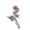
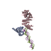
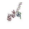
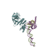
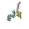
| Title | Structure of Human PARP-1 bound to a DNA double strand break in complex with (2Z)-2-(2,4-dihydroxybenzylidene)-3-oxo-2,3-dihydro-1-benzofuran-7-carboxamide | ||||||
 Components Components |
| ||||||
 Keywords Keywords | TRANSFERASE/DNA/TRANSFERASE INHIBITOR / zinc finger / DNA binding / PARP / polymerase / DNA repair / poly(ADP-ribosyl)ation / TRANSFERASE-DNA-TRANSFERASE INHIBITOR complex | ||||||
| Function / homology |  Function and homology information Function and homology informationNAD+-histone H2BS6 serine ADP-ribosyltransferase activity / NAD+-histone H3S10 serine ADP-ribosyltransferase activity / NAD+-histone H2BE35 glutamate ADP-ribosyltransferase activity / positive regulation of myofibroblast differentiation / negative regulation of ATP biosynthetic process / NAD+-protein-tyrosine ADP-ribosyltransferase activity / NAD+-protein-histidine ADP-ribosyltransferase activity / regulation of base-excision repair / regulation of circadian sleep/wake cycle, non-REM sleep / mitochondrial DNA metabolic process ...NAD+-histone H2BS6 serine ADP-ribosyltransferase activity / NAD+-histone H3S10 serine ADP-ribosyltransferase activity / NAD+-histone H2BE35 glutamate ADP-ribosyltransferase activity / positive regulation of myofibroblast differentiation / negative regulation of ATP biosynthetic process / NAD+-protein-tyrosine ADP-ribosyltransferase activity / NAD+-protein-histidine ADP-ribosyltransferase activity / regulation of base-excision repair / regulation of circadian sleep/wake cycle, non-REM sleep / mitochondrial DNA metabolic process / vRNA Synthesis / carbohydrate biosynthetic process / NAD+-protein-serine ADP-ribosyltransferase activity / NAD DNA ADP-ribosyltransferase activity / negative regulation of adipose tissue development / DNA ADP-ribosylation / regulation of oxidative stress-induced neuron intrinsic apoptotic signaling pathway / ATP generation from poly-ADP-D-ribose / replication fork reversal / positive regulation of necroptotic process / signal transduction involved in regulation of gene expression / transcription regulator activator activity / response to aldosterone / HDR through MMEJ (alt-NHEJ) / positive regulation of DNA-templated transcription, elongation / NAD+ ADP-ribosyltransferase / protein auto-ADP-ribosylation / negative regulation of telomere maintenance via telomere lengthening / NAD+-protein-aspartate ADP-ribosyltransferase activity / mitochondrial DNA repair / positive regulation of intracellular estrogen receptor signaling pathway / protein poly-ADP-ribosylation / NAD+-protein-glutamate ADP-ribosyltransferase activity / negative regulation of cGAS/STING signaling pathway / positive regulation of cardiac muscle hypertrophy / NAD+-protein mono-ADP-ribosyltransferase activity / decidualization / cellular response to zinc ion / protein autoprocessing / positive regulation of mitochondrial depolarization / nuclear replication fork / R-SMAD binding / Transferases; Glycosyltransferases; Pentosyltransferases / positive regulation of SMAD protein signal transduction / negative regulation of transcription elongation by RNA polymerase II / macrophage differentiation / POLB-Dependent Long Patch Base Excision Repair / NAD+ poly-ADP-ribosyltransferase activity / SUMOylation of DNA damage response and repair proteins / nucleosome binding / positive regulation of double-strand break repair via homologous recombination / site of DNA damage / protein localization to chromatin / nucleotidyltransferase activity / positive regulation of adipose tissue development / transforming growth factor beta receptor signaling pathway / negative regulation of innate immune response / telomere maintenance / nuclear estrogen receptor binding / protein modification process / response to gamma radiation / mitochondrion organization / Downregulation of SMAD2/3:SMAD4 transcriptional activity / cellular response to nerve growth factor stimulus / protein-DNA complex / positive regulation of protein localization to nucleus / DNA Damage Recognition in GG-NER / NAD binding / cellular response to amyloid-beta / enzyme activator activity / histone deacetylase binding / Dual Incision in GG-NER / cellular response to insulin stimulus / Formation of Incision Complex in GG-NER / cellular response to UV / nuclear envelope / double-strand break repair / regulation of protein localization / site of double-strand break / cellular response to oxidative stress / transcription regulator complex / transcription by RNA polymerase II / damaged DNA binding / RNA polymerase II-specific DNA-binding transcription factor binding / response to ethanol / positive regulation of canonical NF-kappaB signal transduction / chromosome, telomeric region / nuclear body / innate immune response / DNA repair / negative regulation of DNA-templated transcription / apoptotic process / chromatin binding / DNA damage response / ubiquitin protein ligase binding / protein kinase binding / chromatin / nucleolus / enzyme binding / negative regulation of transcription by RNA polymerase II Similarity search - Function | ||||||
| Biological species |  Homo sapiens (human) Homo sapiens (human) | ||||||
| Method |  X-RAY DIFFRACTION / SYNCHROTRON / MOLECULAR REPLACEMENT / Resolution: 3.65 Å X-RAY DIFFRACTION / SYNCHROTRON / MOLECULAR REPLACEMENT / Resolution: 3.65 Å | ||||||
 Authors Authors | Pascal, J.M. / Steffen, J.D. | ||||||
 Citation Citation | Journal: J.Med.Chem. / Year: 2014 Title: Discovery and Structure-Activity Relationship of Novel 2,3-Dihydrobenzofuran-7-carboxamide and 2,3-Dihydrobenzofuran-3(2H)-one-7-carboxamide Derivatives as Poly(ADP-ribose)polymerase-1 Inhibitors. Authors: Patel, M.R. / Bhatt, A. / Steffen, J.D. / Chergui, A. / Murai, J. / Pommier, Y. / Pascal, J.M. / Trombetta, L.D. / Fronczek, F.R. / Talele, T.T. | ||||||
| History |
|
- Structure visualization
| Structure viewer | Molecule: MolmilJmol/JSmol |
|---|
- Downloads & links
-Download
| PDBx/mmCIF format | 4oqa.cif.gz | 622 KB | Display | PDBx/mmCIF format |
|---|---|---|---|---|
| PDB format | pdb4oqa.ent.gz | 512.3 KB | Display | PDB format |
| PDBx/mmJSON format | 4oqa.json.gz | Tree view | PDBx/mmJSON format | |
| Others |  Other downloads Other downloads |
-Validation report
| Arichive directory | https://data.pdbj.org/pub/pdb/validation_reports/oq/4oqaftp://data.pdbj.org/pub/pdb/validation_reports/oq/4oqa | HTTPS FTP |
|---|
-Related structure data
| Related structure data |  4opxC  4oqbC  4dqyS S: Starting model for refinement C: citing same article ( |
|---|---|
| Similar structure data |

-Links
 PDBj
PDBj


- Assembly
| Deposited unit | 
| ||||||||||||||||||||||||||||||||||||||||||||||||||||||||||||||||||||||||||||||||||||||||||||||||||||||||||||||||||||||||||
|---|---|---|---|---|---|---|---|---|---|---|---|---|---|---|---|---|---|---|---|---|---|---|---|---|---|---|---|---|---|---|---|---|---|---|---|---|---|---|---|---|---|---|---|---|---|---|---|---|---|---|---|---|---|---|---|---|---|---|---|---|---|---|---|---|---|---|---|---|---|---|---|---|---|---|---|---|---|---|---|---|---|---|---|---|---|---|---|---|---|---|---|---|---|---|---|---|---|---|---|---|---|---|---|---|---|---|---|---|---|---|---|---|---|---|---|---|---|---|---|---|---|---|---|
| 1 | 
| ||||||||||||||||||||||||||||||||||||||||||||||||||||||||||||||||||||||||||||||||||||||||||||||||||||||||||||||||||||||||||
| 2 | 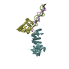
| ||||||||||||||||||||||||||||||||||||||||||||||||||||||||||||||||||||||||||||||||||||||||||||||||||||||||||||||||||||||||||
| Unit cell |
| ||||||||||||||||||||||||||||||||||||||||||||||||||||||||||||||||||||||||||||||||||||||||||||||||||||||||||||||||||||||||||
| Noncrystallographic symmetry (NCS) | NCS domain:
NCS domain segments: Component-ID: _ / Refine code: _
NCS ensembles :
|
-Components
| #1: Protein | Mass: 30520.051 Da / Num. of mol.: 2 / Fragment: N-terminus (Zn1-Zn3, SEE REMARK 999) Source method: isolated from a genetically manipulated source Source: (gene. exp.) Homo sapiens (human) / Gene: PARP1, ADPRT, PPOL / Production host:  #2: Protein | Mass: 57035.020 Da / Num. of mol.: 2 / Fragment: C-terminus (WGR-CAT, UNP residues 518-1014) Source method: isolated from a genetically manipulated source Source: (gene. exp.) Homo sapiens (human) / Gene: PARP1, ADPRT, PPOL / Production host: #3: DNA chain | Mass: 7990.130 Da / Num. of mol.: 2 / Source method: obtained synthetically #4: Chemical | ChemComp-ZN /   Mass: 65.409 Da / Num. of mol.: 4 / Source method: obtained synthetically / Formula: Zn Mass: 65.409 Da / Num. of mol.: 4 / Source method: obtained synthetically / Formula: Zn#5: Chemical | ChemComp-2US / ( |   Mass: 297.262 Da / Num. of mol.: 1 / Source method: obtained synthetically / Formula: C16H11NO5 Mass: 297.262 Da / Num. of mol.: 1 / Source method: obtained synthetically / Formula: C16H11NO5Sequence details | CHAINS A AND D EACH COMPRISE UNP RESIDUES 1-97 AND 207-366 OF PARP-1 CONNECTED BY A ASP-ILE LINKER. | |
|---|
-Experimental details
-Experiment
| Experiment | Method: X-RAY DIFFRACTION / Number of used crystals: 1 |
|---|
- Sample preparation
| Crystal | Density Matthews: 2.87 Å3/Da / Density % sol: 57.07 % |
|---|---|
| Crystal grow | Temperature: 293 K / Method: vapor diffusion, sitting drop / pH: 7.5 Details: 25 mM HEPES, 150 mM sodium chloride, 1 mM EDTA, 0.1 mM TCEP, 6.8% PEG3350, 2% ethylene glycol, pH 7.5, VAPOR DIFFUSION, SITTING DROP, temperature 293K |
-Data collection
| Diffraction | Mean temperature: 100 K |
|---|---|
| Diffraction source | Source: SYNCHROTRON / Site: NSLS  / Beamline: X29A / Wavelength: 1.075 Å / Beamline: X29A / Wavelength: 1.075 Å |
| Detector | Type: ADSC QUANTUM 315r / Detector: CCD / Date: Jul 28, 2013 |
| Radiation | Monochromator: Si(111) / Protocol: SINGLE WAVELENGTH / Monochromatic (M) / Laue (L): M / Scattering type: x-ray |
| Radiation wavelength | Wavelength: 1.075 Å / Relative weight: 1 |
| Reflection | Resolution: 3.65→50 Å / Num. all: 25245 / Num. obs: 24937 |
| Reflection shell | Highest resolution: 3.65 Å |
- Processing
| Software |
| ||||||||||||||||||||||||||||||||||||||||||||||||||||||||||||||||||||||||||||||||||||||||||||||||||||||||||||||||||||||||||||||||||||||||||||||||||||||||||||||||||||||||||||||||||||||
|---|---|---|---|---|---|---|---|---|---|---|---|---|---|---|---|---|---|---|---|---|---|---|---|---|---|---|---|---|---|---|---|---|---|---|---|---|---|---|---|---|---|---|---|---|---|---|---|---|---|---|---|---|---|---|---|---|---|---|---|---|---|---|---|---|---|---|---|---|---|---|---|---|---|---|---|---|---|---|---|---|---|---|---|---|---|---|---|---|---|---|---|---|---|---|---|---|---|---|---|---|---|---|---|---|---|---|---|---|---|---|---|---|---|---|---|---|---|---|---|---|---|---|---|---|---|---|---|---|---|---|---|---|---|---|---|---|---|---|---|---|---|---|---|---|---|---|---|---|---|---|---|---|---|---|---|---|---|---|---|---|---|---|---|---|---|---|---|---|---|---|---|---|---|---|---|---|---|---|---|---|---|---|---|
| Refinement | Method to determine structure: MOLECULAR REPLACEMENT Starting model: PDB ENTRY 4DQY Resolution: 3.65→20 Å / Cor.coef. Fo:Fc: 0.931 / Cor.coef. Fo:Fc free: 0.911 / SU B: 164.771 / SU ML: 0.998 / Cross valid method: THROUGHOUT / ESU R Free: 0.882 / Stereochemistry target values: MAXIMUM LIKELIHOOD / Details: HYDROGENS HAVE BEEN ADDED IN THE RIDING POSITIONS
| ||||||||||||||||||||||||||||||||||||||||||||||||||||||||||||||||||||||||||||||||||||||||||||||||||||||||||||||||||||||||||||||||||||||||||||||||||||||||||||||||||||||||||||||||||||||
| Solvent computation | Ion probe radii: 0.8 Å / Shrinkage radii: 0.8 Å / VDW probe radii: 1.2 Å / Solvent model: MASK | ||||||||||||||||||||||||||||||||||||||||||||||||||||||||||||||||||||||||||||||||||||||||||||||||||||||||||||||||||||||||||||||||||||||||||||||||||||||||||||||||||||||||||||||||||||||
| Displacement parameters | Biso mean: 257.215 Å2
| ||||||||||||||||||||||||||||||||||||||||||||||||||||||||||||||||||||||||||||||||||||||||||||||||||||||||||||||||||||||||||||||||||||||||||||||||||||||||||||||||||||||||||||||||||||||
| Refinement step | Cycle: LAST / Resolution: 3.65→20 Å
| ||||||||||||||||||||||||||||||||||||||||||||||||||||||||||||||||||||||||||||||||||||||||||||||||||||||||||||||||||||||||||||||||||||||||||||||||||||||||||||||||||||||||||||||||||||||
| Refine LS restraints |
|