 Movie
Movie Controller

[English] 日本語
 Yorodumi
- PDB-5euc: The role of the C-terminal region on the oligomeric state and enz... -
+ Open data

- Basic information
| Entry | Database: PDB / ID: 5euc | ||||||
|---|---|---|---|---|---|---|---|
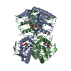
| Title | The role of the C-terminal region on the oligomeric state and enzymatic activity of Trypanosoma cruzi hypoxanthine phosphoribosyl transferase | ||||||
 Components Components | Hypoxanthine-guanine phosphoribosyltransferase | ||||||
 Keywords Keywords | TRANSFERASE / HPRT / phosphoribosyltransferase / T. cruzi / quaternary structure / enzymatic activity modulation / stability / proteolysis / reversible oligomerization / disorder C-terminal region / bisphosphonates | ||||||
| Function / homology |  Function and homology information Function and homology informationhypoxanthine phosphoribosyltransferase / guanine salvage / hypoxanthine metabolic process / hypoxanthine phosphoribosyltransferase activity / GMP salvage / IMP salvage / purine ribonucleoside salvage / nucleotide binding / magnesium ion binding / cytosol Similarity search - Function | ||||||
| Biological species |  | ||||||
| Method |  X-RAY DIFFRACTION / SYNCHROTRON / MOLECULAR REPLACEMENT / Resolution: 2.65 Å X-RAY DIFFRACTION / SYNCHROTRON / MOLECULAR REPLACEMENT / Resolution: 2.65 Å | ||||||
 Authors Authors | Valsecchi, W.M. / Cousido-Siah, A. / Mitschler, A. / Podjarny, A. / Delfino, J.M. / Santos, J. | ||||||
 Citation Citation | Journal: Biochim.Biophys.Acta / Year: 2016 Title: The role of the C-terminal region on the oligomeric state and enzymatic activity of Trypanosoma cruzi hypoxanthine phosphoribosyl transferase. Authors: Valsecchi, W.M. / Cousido-Siah, A. / Defelipe, L.A. / Mitschler, A. / Podjarny, A. / Santos, J. / Delfino, J.M. | ||||||
| History |
|
- Structure visualization
| Structure viewer | Molecule: MolmilJmol/JSmol |
|---|
- Downloads & links
-Download
| PDBx/mmCIF format | 5euc.cif.gz | 166.8 KB | Display | PDBx/mmCIF format |
|---|---|---|---|---|
| PDB format | pdb5euc.ent.gz | 132.6 KB | Display | PDB format |
| PDBx/mmJSON format | 5euc.json.gz | Tree view | PDBx/mmJSON format | |
| Others |  Other downloads Other downloads |
-Validation report
| Arichive directory | https://data.pdbj.org/pub/pdb/validation_reports/eu/5eucftp://data.pdbj.org/pub/pdb/validation_reports/eu/5euc | HTTPS FTP |
|---|
-Related structure data
| Related structure data | 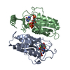 1tc2S S: Starting model for refinement |
|---|---|
| Similar structure data |


-Links
 PDBj
PDBj
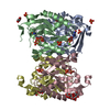
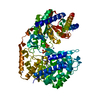
- Assembly
| Deposited unit | 
| ||||||||
|---|---|---|---|---|---|---|---|---|---|
| 1 |
| ||||||||
| Unit cell |
|
-Components
| #1: Protein | Mass: 26666.559 Da / Num. of mol.: 4 / Fragment: UNP residues 21-241 Source method: isolated from a genetically manipulated source Source: (gene. exp.) Strain: CL Brener / Gene: Tc00.1047053509693.70 / Production host:  #2: Water | ChemComp-HOH / |  Mass: 18.015 Da / Num. of mol.: 111 / Source method: isolated from a natural source / Formula: H2O Mass: 18.015 Da / Num. of mol.: 111 / Source method: isolated from a natural source / Formula: H2O |
|---|
-Experimental details
-Experiment
| Experiment | Method: X-RAY DIFFRACTION / Number of used crystals: 1 |
|---|
- Sample preparation
| Crystal | Density Matthews: 1.87 Å3/Da / Density % sol: 34.39 % |
|---|---|
| Crystal grow | Temperature: 293 K / Method: vapor diffusion, sitting drop / pH: 6.5 Details: The drop was a 1:1 mix of protein (in tris 20 mM pH8, NaCl 100 mM) and buffer (1.0 mM MES buffer pH 6.5 and 12 % W/V of PEG 20000) |
-Data collection
| Diffraction | Mean temperature: 100 K |
|---|---|
| Diffraction source | Source: SYNCHROTRON / Site: SLS  / Beamline: X06DA / Wavelength: 0.8 Å / Beamline: X06DA / Wavelength: 0.8 Å |
| Detector | Type: DECTRIS PILATUS 2M / Detector: PIXEL / Date: Dec 2, 2012 |
| Radiation | Monochromator: Bartels Monochromator / Protocol: SINGLE WAVELENGTH / Monochromatic (M) / Laue (L): M / Scattering type: x-ray |
| Radiation wavelength | Wavelength: 0.8 Å / Relative weight: 1 |
| Reflection | Resolution: 2.65→36.34 Å / Num. all: 22566 / Num. obs: 22531 / % possible obs: 99.9 % / Redundancy: 3.7 % / Net I/σ(I): 13.6 |
| Reflection shell | Resolution: 2.65→2.74 Å / Redundancy: 3.7 % / Mean I/σ(I) obs: 3.1 / % possible all: 99.7 |
- Processing
| Software |
| ||||||||||||||||||||||||||||||||||||||||||||||||||||||||||||
|---|---|---|---|---|---|---|---|---|---|---|---|---|---|---|---|---|---|---|---|---|---|---|---|---|---|---|---|---|---|---|---|---|---|---|---|---|---|---|---|---|---|---|---|---|---|---|---|---|---|---|---|---|---|---|---|---|---|---|---|---|---|
| Refinement | Method to determine structure: MOLECULAR REPLACEMENT Starting model: 1TC2 Resolution: 2.65→36.34 Å / Cor.coef. Fo:Fc: 0.942 / Cor.coef. Fo:Fc free: 0.886 / WRfactor Rfree: 0.2491 / WRfactor Rwork: 0.1813 / FOM work R set: 0.8079 / SU B: 13.459 / SU ML: 0.279 / SU Rfree: 0.3889 / Cross valid method: FREE R-VALUE / σ(F): 0 / ESU R Free: 0.389 / Stereochemistry target values: MAXIMUM LIKELIHOOD Details: HYDROGENS HAVE BEEN ADDED IN THE RIDING POSITIONS U VALUES : REFINED INDIVIDUALLY
| ||||||||||||||||||||||||||||||||||||||||||||||||||||||||||||
| Solvent computation | Ion probe radii: 0.8 Å / Shrinkage radii: 0.8 Å / VDW probe radii: 1.2 Å / Solvent model: MASK | ||||||||||||||||||||||||||||||||||||||||||||||||||||||||||||
| Displacement parameters | Biso max: 142.72 Å2 / Biso mean: 44.54 Å2 / Biso min: 21.13 Å2
| ||||||||||||||||||||||||||||||||||||||||||||||||||||||||||||
| Refinement step | Cycle: final / Resolution: 2.65→36.34 Å
| ||||||||||||||||||||||||||||||||||||||||||||||||||||||||||||
| Refine LS restraints |
| ||||||||||||||||||||||||||||||||||||||||||||||||||||||||||||
| LS refinement shell | Resolution: 2.648→2.717 Å / Total num. of bins used: 20
|
