 Movie
Movie Controller

[English] 日本語
 Yorodumi
- PDB-5emy: Human Pancreatic Alpha-Amylase in complex with the mechanism base... -
+ Open data

- Basic information
| Entry | Database: PDB / ID: 5emy | ||||||||||||
|---|---|---|---|---|---|---|---|---|---|---|---|---|---|
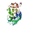
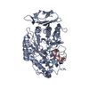
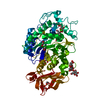
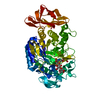
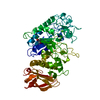
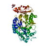
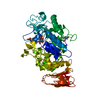
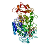
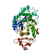
| Title | Human Pancreatic Alpha-Amylase in complex with the mechanism based inactivator glucosyl epi-cyclophellitol | ||||||||||||
 Components Components | Pancreatic alpha-amylase | ||||||||||||
 Keywords Keywords | HYDROLASE/HYDROLASE inhibitor / Amylase / Diabetes / Obesity / Glucosyl hydrolase / HYDROLASE-HYDROLASE inhibitor complex | ||||||||||||
| Function / homology |  Function and homology information Function and homology informationpolysaccharide digestion / Digestion of dietary carbohydrate / Developmental Lineage of Pancreatic Acinar Cells / alpha-amylase / alpha-amylase activity / carbohydrate catabolic process / chloride ion binding / carbohydrate metabolic process / calcium ion binding / : ...polysaccharide digestion / Digestion of dietary carbohydrate / Developmental Lineage of Pancreatic Acinar Cells / alpha-amylase / alpha-amylase activity / carbohydrate catabolic process / chloride ion binding / carbohydrate metabolic process / calcium ion binding / : / extracellular exosome / extracellular region Similarity search - Function | ||||||||||||
| Biological species |  Homo sapiens (human) Homo sapiens (human) | ||||||||||||
| Method |  X-RAY DIFFRACTION / SYNCHROTRON / MOLECULAR REPLACEMENT / Resolution: 1.231 Å X-RAY DIFFRACTION / SYNCHROTRON / MOLECULAR REPLACEMENT / Resolution: 1.231 Å | ||||||||||||
 Authors Authors | Caner, S. / Brayer, G.D. | ||||||||||||
| Funding support |  Canada, 1items Canada, 1items
| ||||||||||||
 Citation Citation | Journal: Febs Lett. / Year: 2016 Title: Glucosyl epi-cyclophellitol allows mechanism-based inactivation and structural analysis of human pancreatic alpha-amylase. Authors: Caner, S. / Zhang, X. / Jiang, J. / Chen, H.M. / Nguyen, N.T. / Overkleeft, H. / Brayer, G.D. / Withers, S.G. | ||||||||||||
| History |
|
- Structure visualization
| Structure viewer | Molecule: MolmilJmol/JSmol |
|---|
- Downloads & links
-Download
| PDBx/mmCIF format | 5emy.cif.gz | 313.4 KB | Display | PDBx/mmCIF format |
|---|---|---|---|---|
| PDB format | pdb5emy.ent.gz | 256.5 KB | Display | PDB format |
| PDBx/mmJSON format | 5emy.json.gz | Tree view | PDBx/mmJSON format | |
| Others |  Other downloads Other downloads |
-Validation report
| Arichive directory | https://data.pdbj.org/pub/pdb/validation_reports/em/5emyftp://data.pdbj.org/pub/pdb/validation_reports/em/5emy | HTTPS FTP |
|---|
-Related structure data
| Related structure data | 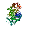 4x9yS S: Starting model for refinement |
|---|---|
| Similar structure data |


-Links
 PDBj
PDBj

- Assembly
| Deposited unit | 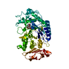
| ||||||||
|---|---|---|---|---|---|---|---|---|---|
| 1 |
| ||||||||
| Unit cell |
|
-Components
| #1: Protein | Mass: 55931.305 Da / Num. of mol.: 1 / Fragment: UNP residues 16-511 Source method: isolated from a genetically manipulated source Details: The residue (XEP) is a modified Aspartate with the complexed Glucosyl-epi-cyclophellitol Source: (gene. exp.) Homo sapiens (human) / Gene: AMY2A / Organ: Pancreas / Production host:  Komagataella pastoris (fungus) / References: UniProt: P04746, alpha-amylase Komagataella pastoris (fungus) / References: UniProt: P04746, alpha-amylase |
|---|---|
| #2: Sugar | ChemComp-5QP / (  Type: D-saccharide / Mass: 340.324 Da / Num. of mol.: 1 / Source method: obtained synthetically / Formula: C13H24O10 Type: D-saccharide / Mass: 340.324 Da / Num. of mol.: 1 / Source method: obtained synthetically / Formula: C13H24O10 |
| #3: Chemical | ChemComp-CL /   Mass: 35.453 Da / Num. of mol.: 1 / Source method: obtained synthetically / Formula: Cl Mass: 35.453 Da / Num. of mol.: 1 / Source method: obtained synthetically / Formula: Cl |
| #4: Chemical | ChemComp-CA /   Mass: 40.078 Da / Num. of mol.: 1 / Source method: obtained synthetically / Formula: Ca Mass: 40.078 Da / Num. of mol.: 1 / Source method: obtained synthetically / Formula: Ca |
| #5: Water | ChemComp-HOH /  Mass: 18.015 Da / Num. of mol.: 559 / Source method: isolated from a natural source / Formula: H2O Mass: 18.015 Da / Num. of mol.: 559 / Source method: isolated from a natural source / Formula: H2O |
| Has protein modification | Y |
| Nonpolymer details | The authors state that the starting material of the ligand is Glucosyl-epi-cyclophellitol |
-Experimental details
-Experiment
| Experiment | Method: X-RAY DIFFRACTION / Number of used crystals: 1 |
|---|
- Sample preparation
| Crystal | Density Matthews: 2.08 Å3/Da / Density % sol: 40.98 % |
|---|---|
| Crystal grow | Temperature: 293 K / Method: vapor diffusion, hanging drop / pH: 7.5 Details: 100 mM sodium cacodylate, 58% MPD, obtained crystals were soaked in 100mM glucosyl epi-cyclophellitol solution and incubated for up to two weeks to allow complex formation. |
-Data collection
| Diffraction | Mean temperature: 100 K |
|---|---|
| Diffraction source | Source: SYNCHROTRON / Site: SSRL  / Beamline: BL12-2 / Wavelength: 0.97946 Å / Beamline: BL12-2 / Wavelength: 0.97946 Å |
| Detector | Type: DECTRIS PILATUS 6M / Detector: PIXEL / Date: Feb 16, 2014 |
| Radiation | Protocol: SINGLE WAVELENGTH / Monochromatic (M) / Laue (L): M / Scattering type: x-ray |
| Radiation wavelength | Wavelength: 0.97946 Å / Relative weight: 1 |
| Reflection | Resolution: 1.23→36.65 Å / Num. all: 134773 / Num. obs: 134741 / % possible obs: 99.5 % / Observed criterion σ(I): 2 / Redundancy: 5.4 % / Rmerge(I) obs: 0.05 / Net I/σ(I): 19.54 |
| Reflection shell | Resolution: 1.23→1.26 Å / Redundancy: 3.7 % / Rmerge(I) obs: 0.75 / Mean I/σ(I) obs: 2.02 / % possible all: 94 |
- Processing
| Software |
| |||||||||||||||||||||||||||||||||||||||||||||||||||||||||||||||||||||||||||||||||||||||||||||||||||||||||||||||||||||||||||||||||||||||||||||||||||||||||||||||||||||||||||||||||||||||||||||||||||||||||||||||||||||||||
|---|---|---|---|---|---|---|---|---|---|---|---|---|---|---|---|---|---|---|---|---|---|---|---|---|---|---|---|---|---|---|---|---|---|---|---|---|---|---|---|---|---|---|---|---|---|---|---|---|---|---|---|---|---|---|---|---|---|---|---|---|---|---|---|---|---|---|---|---|---|---|---|---|---|---|---|---|---|---|---|---|---|---|---|---|---|---|---|---|---|---|---|---|---|---|---|---|---|---|---|---|---|---|---|---|---|---|---|---|---|---|---|---|---|---|---|---|---|---|---|---|---|---|---|---|---|---|---|---|---|---|---|---|---|---|---|---|---|---|---|---|---|---|---|---|---|---|---|---|---|---|---|---|---|---|---|---|---|---|---|---|---|---|---|---|---|---|---|---|---|---|---|---|---|---|---|---|---|---|---|---|---|---|---|---|---|---|---|---|---|---|---|---|---|---|---|---|---|---|---|---|---|---|---|---|---|---|---|---|---|---|---|---|---|---|---|---|---|---|
| Refinement | Method to determine structure: MOLECULAR REPLACEMENT Starting model: 4X9Y Resolution: 1.231→36.648 Å / SU ML: 0.1 / Cross valid method: FREE R-VALUE / σ(F): 1.35 / Phase error: 12.52 / Stereochemistry target values: ML
| |||||||||||||||||||||||||||||||||||||||||||||||||||||||||||||||||||||||||||||||||||||||||||||||||||||||||||||||||||||||||||||||||||||||||||||||||||||||||||||||||||||||||||||||||||||||||||||||||||||||||||||||||||||||||
| Solvent computation | Shrinkage radii: 0.9 Å / VDW probe radii: 1.11 Å / Solvent model: FLAT BULK SOLVENT MODEL | |||||||||||||||||||||||||||||||||||||||||||||||||||||||||||||||||||||||||||||||||||||||||||||||||||||||||||||||||||||||||||||||||||||||||||||||||||||||||||||||||||||||||||||||||||||||||||||||||||||||||||||||||||||||||
| Refinement step | Cycle: LAST / Resolution: 1.231→36.648 Å
| |||||||||||||||||||||||||||||||||||||||||||||||||||||||||||||||||||||||||||||||||||||||||||||||||||||||||||||||||||||||||||||||||||||||||||||||||||||||||||||||||||||||||||||||||||||||||||||||||||||||||||||||||||||||||
| Refine LS restraints |
| |||||||||||||||||||||||||||||||||||||||||||||||||||||||||||||||||||||||||||||||||||||||||||||||||||||||||||||||||||||||||||||||||||||||||||||||||||||||||||||||||||||||||||||||||||||||||||||||||||||||||||||||||||||||||
| LS refinement shell |
|
