 Movie
Movie Controller

[English] 日本語
 Yorodumi
- PDB-1wo2: Crystal structure of the pig pancreatic alpha-amylase complexed w... -
+ Open data

- Basic information
| Entry | Database: PDB / ID: 1wo2 | ||||||||||||
|---|---|---|---|---|---|---|---|---|---|---|---|---|---|
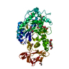
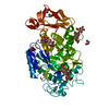
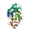
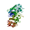
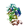
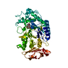
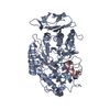
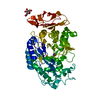
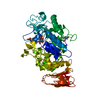
| Title | Crystal structure of the pig pancreatic alpha-amylase complexed with malto-oligosaacharides under the effect of the chloride ion | ||||||||||||
 Components Components | Alpha-amylase, pancreatic | ||||||||||||
 Keywords Keywords | HYDROLASE / BETA-ALPHA-BARRELS | ||||||||||||
| Function / homology |  Function and homology information Function and homology informationalpha-amylase / alpha-amylase activity / carbohydrate catabolic process / chloride ion binding / carbohydrate metabolic process / calcium ion binding / : Similarity search - Function | ||||||||||||
| Biological species |  | ||||||||||||
| Method |  X-RAY DIFFRACTION / MOLECULAR REPLACEMENT / Resolution: 2.01 Å X-RAY DIFFRACTION / MOLECULAR REPLACEMENT / Resolution: 2.01 Å | ||||||||||||
 Authors Authors | Qian, M. / Payan, F. / Nahoum, V. | ||||||||||||
 Citation Citation | Journal: Biochemistry / Year: 2005 Title: Molecular Basis of the Effects of Chloride Ion on the Acid-Base Catalyst in the Mechanism of Pancreatic alpha-Amylase Authors: Qian, M. / Ajandouz, E.H. / Payan, F. / Nahoum, V. #1: Journal: Biochemistry / Year: 2001Title: Enzyme-catalyzed condensation Reaction in a Mammalian alpha-amylase. High-resolution structural analysis of an enzyme-inhibitor complex Authors: Qian, M. / Nahoum, V. / Bonicel, J. / Bischoff, H. / Henrissat, B. / Payan, F. #2: Journal: J.PROTEIN CHEM. / Year: 2003Title: Crystal structure of the pig pancreatic alpha-amylase complexed with malto-oligosaccharides Authors: Payan, F. / Qian, M. | ||||||||||||
| History |
|
- Structure visualization
| Structure viewer | Molecule: MolmilJmol/JSmol |
|---|
- Downloads & links
-Download
| PDBx/mmCIF format | 1wo2.cif.gz | 132.8 KB | Display | PDBx/mmCIF format |
|---|---|---|---|---|
| PDB format | pdb1wo2.ent.gz | 100.4 KB | Display | PDB format |
| PDBx/mmJSON format | 1wo2.json.gz | Tree view | PDBx/mmJSON format | |
| Others |  Other downloads Other downloads |
-Validation report
| Arichive directory | https://data.pdbj.org/pub/pdb/validation_reports/wo/1wo2ftp://data.pdbj.org/pub/pdb/validation_reports/wo/1wo2 | HTTPS FTP |
|---|
-Related structure data
| Related structure data | |
|---|---|
| Similar structure data |


-Links
 PDBj
PDBj

- Assembly
| Deposited unit | 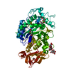
| ||||||||
|---|---|---|---|---|---|---|---|---|---|
| 1 |
| ||||||||
| Unit cell |
|
-Components
-Protein , 1 types, 1 molecules A
| #1: Protein | Mass: 55472.828 Da / Num. of mol.: 1 / Mutation: Q16PCA Source method: isolated from a genetically manipulated source Source: (gene. exp.) Sus scrofa / Tissue: PANCREAS / Production host:  |
|---|
-Sugars , 2 types, 3 molecules
| #2: Polysaccharide | 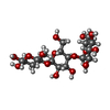  Source method: isolated from a genetically manipulated source Details: oligosaccharide / References: beta-maltotriose #3: Polysaccharide | alpha-D-glucopyranose-(1-4)-beta-D-glucopyranose / beta-maltose | 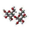  Source method: isolated from a genetically manipulated source Details: oligosaccharide / References: beta-maltose |
|---|
-Non-polymers , 4 types, 757 molecules 


| #4: Chemical | ChemComp-CL /  Mass: 35.453 Da / Num. of mol.: 1 / Source method: obtained synthetically / Formula: Cl Mass: 35.453 Da / Num. of mol.: 1 / Source method: obtained synthetically / Formula: Cl | ||
|---|---|---|---|
| #5: Chemical | ChemComp-CA /  Mass: 40.078 Da / Num. of mol.: 1 / Source method: obtained synthetically / Formula: Ca Mass: 40.078 Da / Num. of mol.: 1 / Source method: obtained synthetically / Formula: Ca | ||
| #6: Chemical | ChemComp-EDO /  Mass: 62.068 Da / Num. of mol.: 6 / Source method: obtained synthetically / Formula: C2H6O2 Mass: 62.068 Da / Num. of mol.: 6 / Source method: obtained synthetically / Formula: C2H6O2#7: Water | ChemComp-HOH / | Mass: 18.015 Da / Num. of mol.: 749 / Source method: isolated from a natural source / Formula: H2O |
-Details
| Has protein modification | Y |
|---|---|
| Sequence details | THESE CONFLICTS ARE EXPLAINED IN REFERENCE AS FOLLOWING, M.QIAN,R.HASER,F.PAYAN STRUCTURE AND ...THESE CONFLICTS ARE EXPLAINED IN REFERENCE AS FOLLOWING, M.QIAN,R.HASER,F.PAYAN STRUCTURE AND MOLECULAR MODEL REFINEMENT |
-Experimental details
-Experiment
| Experiment | Method: X-RAY DIFFRACTION / Number of used crystals: 1 |
|---|
- Sample preparation
| Crystal | Density Matthews: 4.23 Å3/Da / Density % sol: 70 % |
|---|---|
| Crystal grow | Temperature: 277 K / Method: evaporation / pH: 8 Details: Tris, Calcium chloride, Sodium chloride, pH 8, EVAPORATION, temperature 277K |
-Data collection
| Diffraction | Mean temperature: 100 K |
|---|---|
| Diffraction source | Source: ROTATING ANODE / Type: RIGAKU RU300 / Wavelength: 1.5418 Å |
| Detector | Type: MARRESEARCH / Detector: IMAGE PLATE / Date: Oct 15, 2000 |
| Radiation | Monochromator: GRAPHITE / Protocol: SINGLE WAVELENGTH / Monochromatic (M) / Laue (L): M / Scattering type: x-ray |
| Radiation wavelength | Wavelength: 1.5418 Å / Relative weight: 1 |
| Reflection | Resolution: 2.01→30 Å / Num. all: 62412 / Num. obs: 61363 / % possible obs: 98.32 % / Observed criterion σ(F): 1 / Observed criterion σ(I): 1 / Redundancy: 3.7 % / Biso Wilson estimate: 15.3 Å2 / Rmerge(I) obs: 0.067 |
| Reflection shell | Resolution: 2.02→2.07 Å / Redundancy: 3 % / Rmerge(I) obs: 0.173 / Num. unique all: 4603 / % possible all: 98.6 |
- Processing
| Software |
| |||||||||||||||||||||||||
|---|---|---|---|---|---|---|---|---|---|---|---|---|---|---|---|---|---|---|---|---|---|---|---|---|---|---|
| Refinement | Method to determine structure: MOLECULAR REPLACEMENT / Resolution: 2.01→20 Å / Isotropic thermal model: Isotropic / σ(F): 1 / Stereochemistry target values: Engh & Huber
| |||||||||||||||||||||||||
| Refinement step | Cycle: LAST / Resolution: 2.01→20 Å
| |||||||||||||||||||||||||
| Refine LS restraints |
|
