 Movie
Movie Controller

[English] 日本語
 Yorodumi
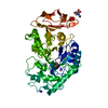
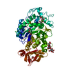
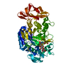
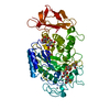
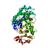
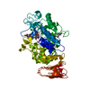
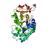
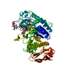
- PDB-1pig: PIG PANCREATIC ALPHA-AMYLASE COMPLEXED WITH THE OLIGOSACCHARIDE V-1532 -
+ Open data

- Basic information
| Entry | Database: PDB / ID: 1pig | ||||||||||||
|---|---|---|---|---|---|---|---|---|---|---|---|---|---|
| Title | PIG PANCREATIC ALPHA-AMYLASE COMPLEXED WITH THE OLIGOSACCHARIDE V-1532 | ||||||||||||
 Components Components | ALPHA-AMYLASE | ||||||||||||
 Keywords Keywords | GLYCOSYLTRANSFERASE / ALPHA-AMYLASE ALPHA-1 / 4-GLUCAN-4-GLUCANOHYDROLASE GLYCOSYLTRANSFERASE | ||||||||||||
| Function / homology |  Function and homology information Function and homology informationalpha-amylase / alpha-amylase activity / carbohydrate catabolic process / chloride ion binding / carbohydrate metabolic process / calcium ion binding / : Similarity search - Function | ||||||||||||
| Biological species |  | ||||||||||||
| Method |  X-RAY DIFFRACTION / DIFFERENCE FOURIER / Resolution: 2.2 Å X-RAY DIFFRACTION / DIFFERENCE FOURIER / Resolution: 2.2 Å | ||||||||||||
 Authors Authors | Machius, M. / Vertesy, L. / Huber, R. / Wiegand, G. | ||||||||||||
 Citation Citation | Journal: J.Mol.Biol. / Year: 1996 Title: Carbohydrate and protein-based inhibitors of porcine pancreatic alpha-amylase: structure analysis and comparison of their binding characteristics. Authors: Machius, M. / Vertesy, L. / Huber, R. / Wiegand, G. #1: Journal: J.Mol.Biol. / Year: 1995Title: The Crystal Structure of Porcine Pancreatic Alpha-Amylase in Complex with the Microbial Inhibitor Tendamistat Authors: Wiegand, G. / Epp, O. / Huber, R. #2: Journal: Protein Sci. / Year: 1995Title: Carbohydrate Binding Sites in a Pancreatic Alpha-Amylase-Substrate Complex, Derived from X-Ray Structure Analysis at 2.1 Angstrom Resolution Authors: Qian, M. / Haser, R. / Payan, F. #3: Journal: J.Mol.Biol. / Year: 1994Title: Refined Molecular Structure of Pig Pancreatic Alpha-Amylase at 2.1 A Resolution Authors: Larson, S.B. / Greenwood, A. / Cascio, D. / Day, J. / McPherson, A. #4: Journal: Biochemistry / Year: 1994Title: The Active Center of a Mammalian Alpha-Amylase. Structure of the Complex of a Pancreatic Alpha-Amylase with a Carbohydrate Inhibitor Refined to 2.2-A Resolution Authors: Qian, M. / Haser, R. / Buisson, G. / Duee, E. / Payan, F. #5: Journal: J.Mol.Biol. / Year: 1993Title: Structure and Molecular Model Refinement of Pig Pancreatic Alpha-Amylase at 2.1 A Resolution Authors: Qian, M. / Haser, R. / Payan, F. | ||||||||||||
| History |
|
- Structure visualization
| Structure viewer | Molecule: MolmilJmol/JSmol |
|---|
- Downloads & links
-Download
| PDBx/mmCIF format | 1pig.cif.gz | 119.8 KB | Display | PDBx/mmCIF format |
|---|---|---|---|---|
| PDB format | pdb1pig.ent.gz | 90.8 KB | Display | PDB format |
| PDBx/mmJSON format | 1pig.json.gz | Tree view | PDBx/mmJSON format | |
| Others |  Other downloads Other downloads |
-Validation report
| Arichive directory | https://data.pdbj.org/pub/pdb/validation_reports/pi/1pigftp://data.pdbj.org/pub/pdb/validation_reports/pi/1pig | HTTPS FTP |
|---|
-Related structure data
| Related structure data | 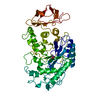 1pifSC S: Starting model for refinement C: citing same article ( |
|---|---|
| Similar structure data |

-Links
 PDBj
PDBj


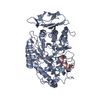
- Assembly
| Deposited unit | 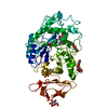
| ||||||||
|---|---|---|---|---|---|---|---|---|---|
| 1 |
| ||||||||
| Unit cell |
|
-Components
-Protein , 1 types, 1 molecules A
| #1: Protein | Mass: 55373.680 Da / Num. of mol.: 1 / Source method: isolated from a natural source / Source: (natural) |
|---|
-Sugars , 5 types, 5 molecules 
| #2: Polysaccharide | 4-amino-4,6-dideoxy-alpha-D-glucopyranose-(1-4)-alpha-D-glucopyranose Source method: isolated from a genetically manipulated source |
|---|---|
| #3: Polysaccharide | 4-amino-4,6-dideoxy-alpha-D-glucopyranose-(1-4)-alpha-D-glucopyranose-(1-4)-beta-D-glucopyranose Source method: isolated from a genetically manipulated source |
| #4: Polysaccharide | alpha-D-glucopyranose-(1-4)-alpha-D-glucopyranose / alpha-maltose  Source method: isolated from a genetically manipulated source Details: oligosaccharide / References: alpha-maltose |
| #5: Polysaccharide | beta-D-glucopyranose-(1-4)-alpha-D-glucopyranose / alpha-cellobiose  Source method: isolated from a genetically manipulated source Details: oligosaccharide / References: alpha-cellobiose |
| #7: Sugar | ChemComp-BGC /  Type: D-saccharide, beta linking / Mass: 180.156 Da / Num. of mol.: 1 Type: D-saccharide, beta linking / Mass: 180.156 Da / Num. of mol.: 1Source method: isolated from a genetically manipulated source Formula: C6H12O6 |
-Non-polymers , 4 types, 231 molecules 


| #6: Chemical | ChemComp-HMC /  Mass: 176.167 Da / Num. of mol.: 1 / Source method: obtained synthetically / Formula: C7H12O5 Mass: 176.167 Da / Num. of mol.: 1 / Source method: obtained synthetically / Formula: C7H12O5 |
|---|---|
| #8: Chemical | ChemComp-CA /  Mass: 40.078 Da / Num. of mol.: 1 / Source method: obtained synthetically / Formula: Ca Mass: 40.078 Da / Num. of mol.: 1 / Source method: obtained synthetically / Formula: Ca |
| #9: Chemical | ChemComp-CL /  Mass: 35.453 Da / Num. of mol.: 1 / Source method: obtained synthetically / Formula: Cl Mass: 35.453 Da / Num. of mol.: 1 / Source method: obtained synthetically / Formula: Cl |
| #10: Water | ChemComp-HOH / Mass: 18.015 Da / Num. of mol.: 228 / Source method: isolated from a natural source / Formula: H2O |
-Details
| Has protein modification | Y |
|---|---|
| Nonpolymer details | TRESTATIN LIKE COMPOUNDS HAVE BEEN ISOLATED FROM STREPTOMYCES GALBUS CULTURE MEDIUM AND TREATED ...TRESTATIN LIKE COMPOUNDS HAVE BEEN ISOLATED FROM STREPTOMYC |
-Experimental details
-Experiment
| Experiment | Method: X-RAY DIFFRACTION / Number of used crystals: 1 |
|---|
- Sample preparation
| Crystal | Density Matthews: 4.39 Å3/Da / Density % sol: 72 % | ||||||||||||||||||||||||||||||
|---|---|---|---|---|---|---|---|---|---|---|---|---|---|---|---|---|---|---|---|---|---|---|---|---|---|---|---|---|---|---|---|
| Crystal grow | Method: vapor diffusion / pH: 6 Details: VAPOR DIFFUSION; 10 MICROLITER OF PROTEIN (15 MG/ML IN 0.1 M AMMONIUM CACODYLATE, PH 10.0 0.001 M V-1532) WERE STEPWISE CONCENTRATED OVER 0.1, 0.15, AND 0.25 M AMMONIUM CACODYLATE, PH 6.0., vapor diffusion PH range: 6.0-10.0 | ||||||||||||||||||||||||||||||
| Crystal grow | *PLUS Method: vapor diffusion | ||||||||||||||||||||||||||||||
| Components of the solutions | *PLUS
|
-Data collection
| Diffraction | Mean temperature: 275 K |
|---|---|
| Diffraction source | Source: ROTATING ANODE / Type: RIGAKU RUH2R / Wavelength: 1.5418 |
| Detector | Type: MARRESEARCH / Detector: IMAGE PLATE / Date: Jul 20, 1995 |
| Radiation | Monochromator: GRAPHITE(002) / Monochromatic (M) / Laue (L): M / Scattering type: x-ray |
| Radiation wavelength | Wavelength: 1.5418 Å / Relative weight: 1 |
| Reflection | Resolution: 2.2→20.5 Å / Num. obs: 47260 / % possible obs: 93.7 % / Observed criterion σ(I): 2 / Redundancy: 4.1 % / Biso Wilson estimate: 32.1 Å2 / Rmerge(I) obs: 0.123 |
| Reflection shell | Resolution: 2.2→2.3 Å / % possible all: 97.8 |
| Reflection shell | *PLUS % possible obs: 97.8 % |
- Processing
| Software |
| ||||||||||||||||||||||||||||||||||||||||||||||||||||||||||||||||||||||||||||||||
|---|---|---|---|---|---|---|---|---|---|---|---|---|---|---|---|---|---|---|---|---|---|---|---|---|---|---|---|---|---|---|---|---|---|---|---|---|---|---|---|---|---|---|---|---|---|---|---|---|---|---|---|---|---|---|---|---|---|---|---|---|---|---|---|---|---|---|---|---|---|---|---|---|---|---|---|---|---|---|---|---|---|
| Refinement | Method to determine structure: DIFFERENCE FOURIER Starting model: 1PIF Resolution: 2.2→7 Å / σ(F): 2
| ||||||||||||||||||||||||||||||||||||||||||||||||||||||||||||||||||||||||||||||||
| Displacement parameters | Biso mean: 28.3 Å2 | ||||||||||||||||||||||||||||||||||||||||||||||||||||||||||||||||||||||||||||||||
| Refine analyze | Luzzati coordinate error obs: 0.2 Å | ||||||||||||||||||||||||||||||||||||||||||||||||||||||||||||||||||||||||||||||||
| Refinement step | Cycle: LAST / Resolution: 2.2→7 Å
| ||||||||||||||||||||||||||||||||||||||||||||||||||||||||||||||||||||||||||||||||
| Refine LS restraints |
| ||||||||||||||||||||||||||||||||||||||||||||||||||||||||||||||||||||||||||||||||
| LS refinement shell | Resolution: 2.2→2.3 Å
| ||||||||||||||||||||||||||||||||||||||||||||||||||||||||||||||||||||||||||||||||
| Software | *PLUS Name: X-PLOR / Classification: refinement | ||||||||||||||||||||||||||||||||||||||||||||||||||||||||||||||||||||||||||||||||
| Refinement | *PLUS | ||||||||||||||||||||||||||||||||||||||||||||||||||||||||||||||||||||||||||||||||
| Solvent computation | *PLUS | ||||||||||||||||||||||||||||||||||||||||||||||||||||||||||||||||||||||||||||||||
| Displacement parameters | *PLUS | ||||||||||||||||||||||||||||||||||||||||||||||||||||||||||||||||||||||||||||||||
| Refine LS restraints | *PLUS
|
