 Movie
Movie Controller

[English] 日本語
 Yorodumi
- PDB-1ua3: Crystal structure of the pig pancreatic a-amylase complexed with ... -
+ Open data

- Basic information
| Entry | Database: PDB / ID: 1ua3 | |||||||||
|---|---|---|---|---|---|---|---|---|---|---|
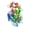
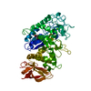
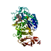
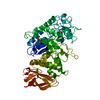
| Title | Crystal structure of the pig pancreatic a-amylase complexed with malto-oligosaccharides | |||||||||
 Components Components | Alpha-amylase, pancreatic | |||||||||
 Keywords Keywords | HYDROLASE / beta-alpha-barrels | |||||||||
| Function / homology |  Function and homology information Function and homology informationalpha-amylase / alpha-amylase activity / carbohydrate catabolic process / chloride ion binding / carbohydrate metabolic process / calcium ion binding / : Similarity search - Function | |||||||||
| Biological species |  | |||||||||
| Method |  X-RAY DIFFRACTION / MOLECULAR REPLACEMENT / Resolution: 2.01 Å X-RAY DIFFRACTION / MOLECULAR REPLACEMENT / Resolution: 2.01 Å | |||||||||
 Authors Authors | Payan, F. / Qian, M. | |||||||||
 Citation Citation | Journal: J.PROTEIN CHEM. / Year: 2003 Title: Crystal Structure of the Pig Pancreatic alpha-Amylase Complexed with Malto-Oligosaccharides Authors: Payan, F. / Qian, M. #1: Journal: PROTEIN SCI. / Year: 1997Title: Structure of a pancreatic alpha-amylase bound to a substrate analogue at 2.03 A resolution Authors: Qian, M. / Spinelli, S. / Driguez, H. / Payan, F. | |||||||||
| History |
|
- Structure visualization
| Structure viewer | Molecule: MolmilJmol/JSmol |
|---|
- Downloads & links
-Download
| PDBx/mmCIF format | 1ua3.cif.gz | 132.2 KB | Display | PDBx/mmCIF format |
|---|---|---|---|---|
| PDB format | pdb1ua3.ent.gz | 100.8 KB | Display | PDB format |
| PDBx/mmJSON format | 1ua3.json.gz | Tree view | PDBx/mmJSON format | |
| Others |  Other downloads Other downloads |
-Validation report
| Arichive directory | https://data.pdbj.org/pub/pdb/validation_reports/ua/1ua3ftp://data.pdbj.org/pub/pdb/validation_reports/ua/1ua3 | HTTPS FTP |
|---|
-Related structure data
| Related structure data | |
|---|---|
| Similar structure data |


-Links
 PDBj
PDBj


- Assembly
| Deposited unit | 
| ||||||||
|---|---|---|---|---|---|---|---|---|---|
| 1 |
| ||||||||
| Unit cell |
|
-Components
-Protein , 1 types, 1 molecules A
| #1: Protein | Mass: 55489.859 Da / Num. of mol.: 1 / Source method: isolated from a natural source / Source: (natural) |
|---|
-Sugars , 3 types, 4 molecules 
| #2: Polysaccharide | alpha-D-glucopyranose-(1-4)-alpha-D-glucopyranose / alpha-maltose  Source method: isolated from a genetically manipulated source Details: oligosaccharide / References: alpha-maltose | ||
|---|---|---|---|
| #3: Polysaccharide |   Source method: isolated from a genetically manipulated source Details: oligosaccharide / References: alpha-maltotriose #6: Sugar | ChemComp-BGC / |  Type: D-saccharide, beta linking / Mass: 180.156 Da / Num. of mol.: 1 Type: D-saccharide, beta linking / Mass: 180.156 Da / Num. of mol.: 1Source method: isolated from a genetically manipulated source Formula: C6H12O6 |
-Non-polymers , 4 types, 768 molecules 


| #4: Chemical |  Mass: 35.453 Da / Num. of mol.: 2 / Source method: obtained synthetically / Formula: Cl Mass: 35.453 Da / Num. of mol.: 2 / Source method: obtained synthetically / Formula: Cl#5: Chemical | ChemComp-CA / |  Mass: 40.078 Da / Num. of mol.: 1 / Source method: obtained synthetically / Formula: Ca Mass: 40.078 Da / Num. of mol.: 1 / Source method: obtained synthetically / Formula: Ca#7: Chemical | ChemComp-EDO /  Mass: 62.068 Da / Num. of mol.: 6 / Source method: obtained synthetically / Formula: C2H6O2 Mass: 62.068 Da / Num. of mol.: 6 / Source method: obtained synthetically / Formula: C2H6O2#8: Water | ChemComp-HOH / | Mass: 18.015 Da / Num. of mol.: 759 / Source method: isolated from a natural source / Formula: H2O |
|---|
-Details
| Has protein modification | Y |
|---|
-Experimental details
-Experiment
| Experiment | Method: X-RAY DIFFRACTION / Number of used crystals: 1 |
|---|
- Sample preparation
| Crystal | Density Matthews: 3.42 Å3/Da / Density % sol: 63.71 % | |||||||||||||||||||||||||||||||||||
|---|---|---|---|---|---|---|---|---|---|---|---|---|---|---|---|---|---|---|---|---|---|---|---|---|---|---|---|---|---|---|---|---|---|---|---|---|
| Crystal grow | Temperature: 277 K / Method: liquid diffusion / pH: 8 Details: Sodium chloride, Calsium chloride, pH 8.0, LIQUID DIFFUSION, temperature 277K | |||||||||||||||||||||||||||||||||||
| Crystal grow | *PLUS Temperature: 4 ℃ / pH: 6.8 / Method: vapor diffusion, hanging drop / Details: White, J.L., (1982) FEBS Lett., 148, 87. | |||||||||||||||||||||||||||||||||||
| Components of the solutions | *PLUS
|
-Data collection
| Diffraction | Mean temperature: 298 K |
|---|---|
| Diffraction source | Source: ROTATING ANODE / Type: RIGAKU RU300 / Wavelength: 1.5418 Å |
| Detector | Type: MARRESEARCH / Detector: IMAGE PLATE / Date: Apr 20, 1996 |
| Radiation | Monochromator: GRAPHITE / Protocol: SINGLE WAVELENGTH / Monochromatic (M) / Laue (L): M / Scattering type: x-ray |
| Radiation wavelength | Wavelength: 1.5418 Å / Relative weight: 1 |
| Reflection | Resolution: 2.01→30 Å / Num. all: 62336 / Num. obs: 61298 / % possible obs: 98.3 % / Observed criterion σ(F): 1 / Observed criterion σ(I): 1 / Redundancy: 3.7 % / Biso Wilson estimate: 15.3 Å2 / Rmerge(I) obs: 0.055 / Net I/σ(I): 9.5 |
| Reflection shell | Resolution: 2.01→2.04 Å / Redundancy: 3.6 % / Rmerge(I) obs: 0.218 / Mean I/σ(I) obs: 1.6 / % possible all: 85.8 |
| Reflection | *PLUS Lowest resolution: 35 Å / Num. obs: 61415 / Num. measured all: 211556 |
| Reflection shell | *PLUS % possible obs: 85.8 % |
- Processing
| Software |
| |||||||||||||||||||||||||
|---|---|---|---|---|---|---|---|---|---|---|---|---|---|---|---|---|---|---|---|---|---|---|---|---|---|---|
| Refinement | Method to determine structure: MOLECULAR REPLACEMENT / Resolution: 2.01→30 Å / σ(F): 1 / Stereochemistry target values: Engh & Huber
| |||||||||||||||||||||||||
| Refinement step | Cycle: LAST / Resolution: 2.01→30 Å
| |||||||||||||||||||||||||
| Refine LS restraints |
| |||||||||||||||||||||||||
| Refinement | *PLUS Lowest resolution: 35 Å / Rfactor Rfree: 0.198 / Rfactor Rwork: 0.1701 | |||||||||||||||||||||||||
| Solvent computation | *PLUS | |||||||||||||||||||||||||
| Displacement parameters | *PLUS | |||||||||||||||||||||||||
| Refine LS restraints | *PLUS
| |||||||||||||||||||||||||
| LS refinement shell | *PLUS Highest resolution: 2.01 Å / Lowest resolution: 2.08 Å / Rfactor Rfree: 0.2349 / Rfactor Rwork: 0.2182 |
