 Movie
Movie Controller


+ Open data

- Basic information
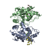
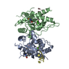
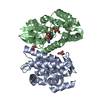
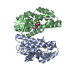
| Entry | Database: PDB / ID: 5ecs | ||||||
|---|---|---|---|---|---|---|---|
| Title | Crystal Structure of FIP1 with GSH | ||||||
 Components Components | Glutathione S-transferase U20 | ||||||
 Keywords Keywords | TRANSFERASE / Glutathione S-transferase | ||||||
| Function / homology |  Function and homology information Function and homology informationregulation of response to red or far red light / toxin catabolic process / glutathione binding / apoplast / response to gravity / glutathione transferase / glutathione transferase activity / chloroplast / glutathione metabolic process / enzyme binding ...regulation of response to red or far red light / toxin catabolic process / glutathione binding / apoplast / response to gravity / glutathione transferase / glutathione transferase activity / chloroplast / glutathione metabolic process / enzyme binding / mitochondrion / nucleus / cytoplasm / cytosol Similarity search - Function | ||||||
| Biological species |  | ||||||
| Method |  X-RAY DIFFRACTION / SYNCHROTRON / MOLECULAR REPLACEMENT / Resolution: 1.65000262305 Å X-RAY DIFFRACTION / SYNCHROTRON / MOLECULAR REPLACEMENT / Resolution: 1.65000262305 Å | ||||||
 Authors Authors | Chen, C.Y. / Cheng, Y.S. | ||||||
 Citation Citation | Journal: Proc. Natl. Acad. Sci. U.S.A. / Year: 2017 Title: Structural basis of jasmonate-amido synthetase FIN219 in complex with glutathione S-transferase FIP1 during the JA signal regulation Authors: Chen, C.Y. / Ho, S.S. / Kuo, T.Y. / Hsieh, H.L. / Cheng, Y.S. | ||||||
| History |
|
- Structure visualization
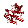
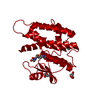
| Structure viewer | Molecule: MolmilJmol/JSmol |
|---|
- Downloads & links
-Download
| PDBx/mmCIF format | 5ecs.cif.gz | 108.5 KB | Display | PDBx/mmCIF format |
|---|---|---|---|---|
| PDB format | pdb5ecs.ent.gz | 81.2 KB | Display | PDB format |
| PDBx/mmJSON format | 5ecs.json.gz | Tree view | PDBx/mmJSON format | |
| Others |  Other downloads Other downloads |
-Validation report
| Arichive directory | https://data.pdbj.org/pub/pdb/validation_reports/ec/5ecsftp://data.pdbj.org/pub/pdb/validation_reports/ec/5ecs | HTTPS FTP |
|---|
-Related structure data
| Related structure data |  5echC  5eciC  5eckC  5eclC  5ecmC  5ecnC  5ecoC  5ecpC  5ecqC  5ecrC  5gzzC 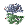 2vo4S C: citing same article ( S: Starting model for refinement |
|---|---|
| Similar structure data |


-Links
 PDBj
PDBj

- Assembly
| Deposited unit | 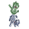
| ||||||||||
|---|---|---|---|---|---|---|---|---|---|---|---|
| 1 | 
| ||||||||||
| 2 | 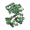
| ||||||||||
| Unit cell |
| ||||||||||
| Components on special symmetry positions |
|
-Components
| #1: Protein | Mass: 25867.312 Da / Num. of mol.: 2 Source method: isolated from a genetically manipulated source Source: (gene. exp.)  #2: Chemical |   Mass: 307.323 Da / Num. of mol.: 2 / Source method: obtained synthetically / Formula: C10H17N3O6S Mass: 307.323 Da / Num. of mol.: 2 / Source method: obtained synthetically / Formula: C10H17N3O6S#3: Water | ChemComp-HOH / |  Mass: 18.015 Da / Num. of mol.: 268 / Source method: isolated from a natural source / Formula: H2O Mass: 18.015 Da / Num. of mol.: 268 / Source method: isolated from a natural source / Formula: H2O |
|---|
-Experimental details
-Experiment
| Experiment | Method: X-RAY DIFFRACTION / Number of used crystals: 1 |
|---|
- Sample preparation
| Crystal | Density Matthews: 2.59 Å3/Da / Density % sol: 52.52 % |
|---|---|
| Crystal grow | Temperature: 297 K / Method: vapor diffusion, sitting drop / pH: 8.5 Details: 0.2 M sodium acetate trihydrate, 0.1 M Tris-HCl, 30%(w/v) PEG 3000 |
-Data collection
| Diffraction | Mean temperature: 100 K |
|---|---|
| Diffraction source | Source: SYNCHROTRON / Site: NSRRC  / Beamline: BL13C1 / Wavelength: 0.97622 Å / Beamline: BL13C1 / Wavelength: 0.97622 Å |
| Detector | Type: RAYONIX MX-300 / Detector: CCD / Date: Nov 8, 2012 |
| Radiation | Protocol: SINGLE WAVELENGTH / Monochromatic (M) / Laue (L): M / Scattering type: x-ray |
| Radiation wavelength | Wavelength: 0.97622 Å / Relative weight: 1 |
| Reflection | Resolution: 1.65→50 Å / Num. obs: 60431 / % possible obs: 99 % / Observed criterion σ(F): 0 / Observed criterion σ(I): -3 / Redundancy: 3.5 % / Biso Wilson estimate: 17.4150366692 Å2 / Rmerge(I) obs: 0.054 / Rsym value: 0.054 / Net I/av σ(I): 21.624 / Net I/σ(I): 13.9 |
| Reflection shell | Resolution: 1.65→1.71 Å / Redundancy: 3.4 % / Rmerge(I) obs: 0.358 / Mean I/σ(I) obs: 4.09 / Rsym value: 0.358 / % possible all: 97.1 |
- Processing
| Software |
| |||||||||||||||||||||||||||||||||||||||||||||||||||||||||||||||||||||||||||||||||||||||||||||||||||||||||
|---|---|---|---|---|---|---|---|---|---|---|---|---|---|---|---|---|---|---|---|---|---|---|---|---|---|---|---|---|---|---|---|---|---|---|---|---|---|---|---|---|---|---|---|---|---|---|---|---|---|---|---|---|---|---|---|---|---|---|---|---|---|---|---|---|---|---|---|---|---|---|---|---|---|---|---|---|---|---|---|---|---|---|---|---|---|---|---|---|---|---|---|---|---|---|---|---|---|---|---|---|---|---|---|---|---|---|
| Refinement | Method to determine structure: MOLECULAR REPLACEMENT Starting model: 2VO4 Resolution: 1.65000262305→26.6503929074 Å / SU ML: 0.146807005221 / Cross valid method: FREE R-VALUE / σ(F): 1.34368788552 / Phase error: 18.3123962951
| |||||||||||||||||||||||||||||||||||||||||||||||||||||||||||||||||||||||||||||||||||||||||||||||||||||||||
| Solvent computation | Shrinkage radii: 0.9 Å / VDW probe radii: 1.11 Å / Solvent model: FLAT BULK SOLVENT MODEL | |||||||||||||||||||||||||||||||||||||||||||||||||||||||||||||||||||||||||||||||||||||||||||||||||||||||||
| Displacement parameters | Biso mean: 20.3235357518 Å2 | |||||||||||||||||||||||||||||||||||||||||||||||||||||||||||||||||||||||||||||||||||||||||||||||||||||||||
| Refinement step | Cycle: final / Resolution: 1.65000262305→26.6503929074 Å
| |||||||||||||||||||||||||||||||||||||||||||||||||||||||||||||||||||||||||||||||||||||||||||||||||||||||||
| Refine LS restraints |
| |||||||||||||||||||||||||||||||||||||||||||||||||||||||||||||||||||||||||||||||||||||||||||||||||||||||||
| LS refinement shell |
|
