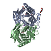
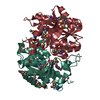
HYDROLASE/DNA / RNase H-like fold / Polyproline type II helix / HYDROLASE-DNA complex
Function / homology
Function and homology information
cellular response to type I interferon / immune response in brain or nervous system / adenyl deoxyribonucleotide binding / immune complex formation / activation of immune response / organ or tissue specific immune response / DNA synthesis involved in UV-damage excision repair / atrial cardiac muscle tissue development / T cell antigen processing and presentation / MutSalpha complex binding ...cellular response to type I interferon / immune response in brain or nervous system / adenyl deoxyribonucleotide binding / immune complex formation / activation of immune response / organ or tissue specific immune response / DNA synthesis involved in UV-damage excision repair / atrial cardiac muscle tissue development / T cell antigen processing and presentation / MutSalpha complex binding / retrotransposition / DNA exonuclease activity / DNA modification / regulation of lipid biosynthetic process / oligosaccharyltransferase complex / regulation of fatty acid metabolic process / heart process / regulation of protein complex stability / exodeoxyribonuclease III / lymphoid progenitor cell differentiation / double-stranded DNA 3'-5' DNA exonuclease activity / regulation of type I interferon production / regulation of lysosome organization / glycoprotein biosynthetic process / cellular response to hydroxyurea / regulation of tumor necrosis factor production / MutLalpha complex binding / 3'-5'-DNA exonuclease activity / regulation of immunoglobulin production / inflammatory response to antigenic stimulus / macrophage activation involved in immune response / regulation of cellular respiration / DNA catabolic process / regulation of T cell activation / apoptotic cell clearance / regulation of glycolytic process / DNA binding, bending / cGAS/STING signaling pathway / negative regulation of type I interferon-mediated signaling pathway / WW domain binding / regulation of innate immune response / DNA metabolic process / negative regulation of cGAS/STING signaling pathway / type I interferon-mediated signaling pathway / blood vessel development / nuclear replication fork / response to UV / heart morphogenesis / cellular response to interferon-beta / mitotic G1 DNA damage checkpoint signaling / 3'-5' exonuclease activity / negative regulation of innate immune response / DNA damage checkpoint signaling / determination of adult lifespan / generation of precursor metabolites and energy / kidney development / cellular response to reactive oxygen species / establishment of protein localization / cellular response to gamma radiation / protein-DNA complex / cellular response to UV / single-stranded DNA binding / regulation of gene expression / cellular response to oxidative stress / regulation of inflammatory response / double-stranded DNA binding / defense response to virus / adaptive immune response / DNA replication / protein stabilization / immune response / inflammatory response / innate immune response / DNA damage response / endoplasmic reticulum membrane / magnesium ion binding / endoplasmic reticulum / protein homodimerization activity / DNA binding / identical protein binding / nucleus / cytoplasm / cytosol Similarity search - Function
In the structure databanks used in Yorodumi, some data are registered as the other names, "COVID-19 virus" and "2019-nCoV". Here are the details of the virus and the list of structure data.
Jan 31, 2019. EMDB accession codes are about to change! (news from PDBe EMDB page)
EMDB accession codes are about to change! (news from PDBe EMDB page)
The allocation of 4 digits for EMDB accession codes will soon come to an end. Whilst these codes will remain in use, new EMDB accession codes will include an additional digit and will expand incrementally as the available range of codes is exhausted. The current 4-digit format prefixed with “EMD-” (i.e. EMD-XXXX) will advance to a 5-digit format (i.e. EMD-XXXXX), and so on. It is currently estimated that the 4-digit codes will be depleted around Spring 2019, at which point the 5-digit format will come into force.
The EM Navigator/Yorodumi systems omit the EMD- prefix.
Related info.:Q: What is EMD? / ID/Accession-code notation in Yorodumi/EM Navigator
Yorodumi is a browser for structure data from EMDB, PDB, SASBDB, etc.
This page is also the successor to EM Navigator detail page, and also detail information page/front-end page for Omokage search.
The word "yorodu" (or yorozu) is an old Japanese word meaning "ten thousand". "mi" (miru) is to see.
Related info.:EMDB / PDB / SASBDB / Comparison of 3 databanks / Yorodumi Search / Aug 31, 2016. New EM Navigator & Yorodumi / Yorodumi Papers / Jmol/JSmol / Function and homology information / Changes in new EM Navigator and Yorodumi
 Movie
Movie Controller


 Open data
 Basic information
 Components
 Keywords
 Function and homology information
 X-RAY DIFFRACTION /
  Authors
 Citation
 Structure visualization
 Downloads & links
 Other downloads
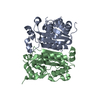


 PDBj
PDBj Assembly


 Mass: 40.078 Da / Num. of mol.: 4 / Source method: obtained synthetically / Formula: Ca
 Mass: 18.015 Da / Num. of mol.: 93 / Source method: isolated from a natural source / Formula: H2O
 Sample preparation
 Processing