 Movie
Movie Controller

[English] 日本語
 Yorodumi
- PDB-5kej: Crystallographic structure of the Tau class glutathione S-transfe... -
+ Open data

- Basic information
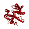
| Entry | Database: PDB / ID: 5kej | ||||||
|---|---|---|---|---|---|---|---|
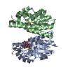
| Title | Crystallographic structure of the Tau class glutathione S-transferase MiGSTU in complex with S-hexyl-glutathione | ||||||
 Components Components | Tau class glutathione S-transferase | ||||||
 Keywords Keywords | TRANSFERASE / detoxification | ||||||
| Function / homology |  Function and homology information Function and homology informationtoxin catabolic process / glutathione transferase / glutathione transferase activity / glutathione metabolic process / cytosol Similarity search - Function | ||||||
| Biological species |  Mangifera indica (mango) Mangifera indica (mango) | ||||||
| Method |  X-RAY DIFFRACTION / SYNCHROTRON / MOLECULAR REPLACEMENT / Resolution: 2.35 Å X-RAY DIFFRACTION / SYNCHROTRON / MOLECULAR REPLACEMENT / Resolution: 2.35 Å | ||||||
 Authors Authors | Valenzuela-Chavira, I. / Serrano-Posada, H. / Lopez-Zavala, A. / Hernandez-Paredes, J. / Sotelo-Mundo, R. | ||||||
 Citation Citation | Journal: Biochimie / Year: 2017 Title: Insights into ligand binding to a glutathione S-transferase from mango: Structure, thermodynamics and kinetics. Authors: Valenzuela-Chavira, I. / Contreras-Vergara, C.A. / Arvizu-Flores, A.A. / Serrano-Posada, H. / Lopez-Zavala, A.A. / Garcia-Orozco, K.D. / Hernandez-Paredes, J. / Rudino-Pinera, E. / ...Authors: Valenzuela-Chavira, I. / Contreras-Vergara, C.A. / Arvizu-Flores, A.A. / Serrano-Posada, H. / Lopez-Zavala, A.A. / Garcia-Orozco, K.D. / Hernandez-Paredes, J. / Rudino-Pinera, E. / Stojanoff, V. / Sotelo-Mundo, R.R. / Islas-Osuna, M.A. | ||||||
| History |
|
- Structure visualization
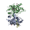
| Structure viewer | Molecule: MolmilJmol/JSmol |
|---|
- Downloads & links
-Download
| PDBx/mmCIF format | 5kej.cif.gz | 105.2 KB | Display | PDBx/mmCIF format |
|---|---|---|---|---|
| PDB format | pdb5kej.ent.gz | 80.1 KB | Display | PDB format |
| PDBx/mmJSON format | 5kej.json.gz | Tree view | PDBx/mmJSON format | |
| Others |  Other downloads Other downloads |
-Validation report
| Arichive directory | https://data.pdbj.org/pub/pdb/validation_reports/ke/5kejftp://data.pdbj.org/pub/pdb/validation_reports/ke/5kej | HTTPS FTP |
|---|
-Related structure data
| Related structure data | 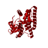 5g5eC 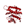 5g5fC  1gwcS S: Starting model for refinement C: citing same article ( |
|---|---|
| Similar structure data |


-Links
 PDBj
PDBj

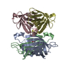
- Assembly
| Deposited unit | 
| ||||||||||
|---|---|---|---|---|---|---|---|---|---|---|---|
| 1 |
| ||||||||||
| Unit cell |
|
-Components
| #1: Protein | Mass: 25550.686 Da / Num. of mol.: 2 Source method: isolated from a genetically manipulated source Source: (gene. exp.) Mangifera indica (mango) / Plasmid: pJExpress404 / Production host:  References: UniProt: A0A1P8NWC2*PLUS, glutathione transferase #2: Chemical | 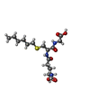  Mass: 392.491 Da / Num. of mol.: 2 / Source method: obtained synthetically / Formula: C16H30N3O6S Mass: 392.491 Da / Num. of mol.: 2 / Source method: obtained synthetically / Formula: C16H30N3O6S#3: Chemical | ChemComp-PEG /   Mass: 106.120 Da / Num. of mol.: 6 / Source method: obtained synthetically / Formula: C4H10O3 Mass: 106.120 Da / Num. of mol.: 6 / Source method: obtained synthetically / Formula: C4H10O3#4: Water | ChemComp-HOH / |  Mass: 18.015 Da / Num. of mol.: 124 / Source method: isolated from a natural source / Formula: H2O Mass: 18.015 Da / Num. of mol.: 124 / Source method: isolated from a natural source / Formula: H2O |
|---|
-Experimental details
-Experiment
| Experiment | Method: X-RAY DIFFRACTION / Number of used crystals: 1 |
|---|
- Sample preparation
| Crystal | Density Matthews: 2.7 Å3/Da / Density % sol: 54.5 % |
|---|---|
| Crystal grow | Temperature: 289 K / Method: batch mode / pH: 6 Details: 0.2 M ammonium acetate, 0.1 M Bis-Tris pH 6.0, 25%(w/v) polyethylene glycol 3350, 5 mM S-hexyl-glutathione |
-Data collection
| Diffraction | Mean temperature: 100 K |
|---|---|
| Diffraction source | Source: SYNCHROTRON / Site: SSRL  / Beamline: BL14-1 / Wavelength: 1.181 Å / Beamline: BL14-1 / Wavelength: 1.181 Å |
| Detector | Type: MARMOSAIC 325 mm CCD / Detector: CCD / Date: May 21, 2016 |
| Radiation | Protocol: SINGLE WAVELENGTH / Monochromatic (M) / Laue (L): M / Scattering type: x-ray |
| Radiation wavelength | Wavelength: 1.181 Å / Relative weight: 1 |
| Reflection | Resolution: 2.35→40.343 Å / Num. obs: 23652 / % possible obs: 100 % / Redundancy: 14.6 % / Biso Wilson estimate: 36.3 Å2 / Rmerge(I) obs: 0.17 / Net I/σ(I): 11.8 |
| Reflection shell | Resolution: 2.35→2.48 Å / Redundancy: 14.7 % / Mean I/σ(I) obs: 3.3 / % possible all: 100 |
- Processing
| Software |
| ||||||||||||||||||||||||||||||||||||||||||||||||||||||
|---|---|---|---|---|---|---|---|---|---|---|---|---|---|---|---|---|---|---|---|---|---|---|---|---|---|---|---|---|---|---|---|---|---|---|---|---|---|---|---|---|---|---|---|---|---|---|---|---|---|---|---|---|---|---|---|
| Refinement | Method to determine structure: MOLECULAR REPLACEMENT Starting model: Homology model based on the structure of wheat Tau class glutathione S-transferase (PDB entry 1GWC). Resolution: 2.35→40.343 Å / SU ML: 0.21 / Cross valid method: FREE R-VALUE / σ(F): 1.34 / Phase error: 24.44
| ||||||||||||||||||||||||||||||||||||||||||||||||||||||
| Solvent computation | Shrinkage radii: 0.9 Å / VDW probe radii: 1.11 Å | ||||||||||||||||||||||||||||||||||||||||||||||||||||||
| Displacement parameters | Biso max: 109.69 Å2 / Biso mean: 43.4792 Å2 / Biso min: 21.14 Å2 | ||||||||||||||||||||||||||||||||||||||||||||||||||||||
| Refinement step | Cycle: final / Resolution: 2.35→40.343 Å
| ||||||||||||||||||||||||||||||||||||||||||||||||||||||
| Refine LS restraints |
| ||||||||||||||||||||||||||||||||||||||||||||||||||||||
| LS refinement shell | Refine-ID: X-RAY DIFFRACTION / Total num. of bins used: 8 / % reflection obs: 100 %
|
