 Movie
Movie Controller


+ Open data

- Basic information
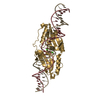
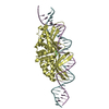
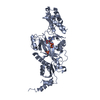
| Entry | Database: PDB / ID: 5e5p | ||||||
|---|---|---|---|---|---|---|---|
| Title | Wild type I-SmaMI in the space group of C121 | ||||||
 Components Components | I-SmaMI LAGLIDADG meganuclease | ||||||
 Keywords Keywords | HYDROLASE / LAGLIDADG / I-SmaMI | ||||||
| Function / homology |  Function and homology information Function and homology information | ||||||
| Biological species |  Sordaria macrospora (fungus) Sordaria macrospora (fungus) | ||||||
| Method |  X-RAY DIFFRACTION / MOLECULAR REPLACEMENT / Resolution: 2.65 Å X-RAY DIFFRACTION / MOLECULAR REPLACEMENT / Resolution: 2.65 Å | ||||||
 Authors Authors | Shen, B.W. | ||||||
| Funding support |  United States, 1items United States, 1items
| ||||||
 Citation Citation | Journal: J.Mol.Biol. / Year: 2016 Title: The Structural Basis of Asymmetry in DNA Binding and Cleavage as Exhibited by the I-SmaMI LAGLIDADG Meganuclease. Authors: Shen, B.W. / Lambert, A. / Walker, B.C. / Stoddard, B.L. / Kaiser, B.K. | ||||||
| History |
|
- Structure visualization
| Structure viewer | Molecule: MolmilJmol/JSmol |
|---|
- Downloads & links
-Download
| PDBx/mmCIF format | 5e5p.cif.gz | 132.6 KB | Display | PDBx/mmCIF format |
|---|---|---|---|---|
| PDB format | pdb5e5p.ent.gz | 103.8 KB | Display | PDB format |
| PDBx/mmJSON format | 5e5p.json.gz | Tree view | PDBx/mmJSON format | |
| Others |  Other downloads Other downloads |
-Validation report
| Summary document | 5e5p_validation.pdf.gz | 454.2 KB | Display | wwPDB validaton report |
|---|---|---|---|---|
| Full document | 5e5p_full_validation.pdf.gz | 455.6 KB | Display | |
| Data in XML | 5e5p_validation.xml.gz | 13.6 KB | Display | |
| Data in CIF | 5e5p_validation.cif.gz | 18.7 KB | Display | |
| Arichive directory | https://data.pdbj.org/pub/pdb/validation_reports/e5/5e5pftp://data.pdbj.org/pub/pdb/validation_reports/e5/5e5p | HTTPS FTP |
-Related structure data
| Related structure data |  5e5oC  5e5sC  5e63C  5e67C 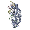 4loxS C: citing same article ( S: Starting model for refinement |
|---|---|
| Similar structure data |


-Links
 PDBj
PDBj

- Assembly
| Deposited unit | 
| ||||||||
|---|---|---|---|---|---|---|---|---|---|
| 1 |
| ||||||||
| Unit cell |
|
-Components
| #1: Protein | Mass: 34152.641 Da / Num. of mol.: 1 / Fragment: UNP residues 114-415 Source method: isolated from a genetically manipulated source Source: (gene. exp.) Sordaria macrospora (strain ATCC MYA-333 / DSM 997 / K(L3346) / K-hell) (fungus)Strain: ATCC MYA-333 / DSM 997 / K(L3346) / K-hell / Gene: SMAC_12671 / Plasmid: pET21d(+) / Production host:  | ||
|---|---|---|---|
| #2: Chemical | ChemComp-PG0 /   Mass: 120.147 Da / Num. of mol.: 1 / Source method: obtained synthetically / Formula: C5H12O3 / Comment: inhibitor, precipitant*YM Mass: 120.147 Da / Num. of mol.: 1 / Source method: obtained synthetically / Formula: C5H12O3 / Comment: inhibitor, precipitant*YM | ||
| #3: Chemical |   Mass: 62.068 Da / Num. of mol.: 2 / Source method: obtained synthetically / Formula: C2H6O2 Mass: 62.068 Da / Num. of mol.: 2 / Source method: obtained synthetically / Formula: C2H6O2#4: Water | ChemComp-HOH / |  Mass: 18.015 Da / Num. of mol.: 84 / Source method: isolated from a natural source / Formula: H2O Mass: 18.015 Da / Num. of mol.: 84 / Source method: isolated from a natural source / Formula: H2O |
-Experimental details
-Experiment
| Experiment | Method: X-RAY DIFFRACTION / Number of used crystals: 1 |
|---|
- Sample preparation
| Crystal | Density Matthews: 2.65 Å3/Da / Density % sol: 53.59 % |
|---|---|
| Crystal grow | Temperature: 298 K / Method: vapor diffusion, sitting drop / pH: 7.5 / Details: 20% PEG8K, HEPES / PH range: 7.5 |
-Data collection
| Diffraction | Mean temperature: 100 K |
|---|---|
| Diffraction source | Source: ROTATING ANODE / Type: RIGAKU MICROMAX-007 HF / Wavelength: 1.54 Å |
| Detector | Type: RIGAKU SATURN 70 / Detector: CCD / Date: Sep 4, 2013 |
| Radiation | Protocol: SINGLE WAVELENGTH / Monochromatic (M) / Laue (L): M / Scattering type: x-ray |
| Radiation wavelength | Wavelength: 1.54 Å / Relative weight: 1 |
| Reflection | Resolution: 2.64→27.1 Å / Num. obs: 10491 / % possible obs: 98.5 % / Observed criterion σ(F): 1 / Observed criterion σ(I): 1 / Redundancy: 3.7 % / Biso Wilson estimate: 21.2 Å2 / Rsym value: 0.05 / Net I/σ(I): 22 |
| Reflection shell | Resolution: 2.64→2.73 Å / Redundancy: 3.1 % / Rmerge(I) obs: 0.149 / Mean I/σ(I) obs: 7.5 / % possible all: 87 |
- Processing
| Software |
| ||||||||||||||||||||||||||||||||||||||||||||||||||||||||||||||||||||||||||||||||||||||||||||||||||||||||||||||||||||||||||||||||||||||||||||||||||||||||||||||||||||||||||||||||||||||
|---|---|---|---|---|---|---|---|---|---|---|---|---|---|---|---|---|---|---|---|---|---|---|---|---|---|---|---|---|---|---|---|---|---|---|---|---|---|---|---|---|---|---|---|---|---|---|---|---|---|---|---|---|---|---|---|---|---|---|---|---|---|---|---|---|---|---|---|---|---|---|---|---|---|---|---|---|---|---|---|---|---|---|---|---|---|---|---|---|---|---|---|---|---|---|---|---|---|---|---|---|---|---|---|---|---|---|---|---|---|---|---|---|---|---|---|---|---|---|---|---|---|---|---|---|---|---|---|---|---|---|---|---|---|---|---|---|---|---|---|---|---|---|---|---|---|---|---|---|---|---|---|---|---|---|---|---|---|---|---|---|---|---|---|---|---|---|---|---|---|---|---|---|---|---|---|---|---|---|---|---|---|---|---|
| Refinement | Method to determine structure: MOLECULAR REPLACEMENT Starting model: 4LOX Resolution: 2.65→27.1 Å / Cor.coef. Fo:Fc: 0.946 / Cor.coef. Fo:Fc free: 0.866 / SU B: 23.617 / SU ML: 0.246 / Cross valid method: THROUGHOUT / ESU R: 3.427 / ESU R Free: 0.357 / Stereochemistry target values: MAXIMUM LIKELIHOOD / Details: HYDROGENS HAVE BEEN ADDED IN THE RIDING POSITIONS
| ||||||||||||||||||||||||||||||||||||||||||||||||||||||||||||||||||||||||||||||||||||||||||||||||||||||||||||||||||||||||||||||||||||||||||||||||||||||||||||||||||||||||||||||||||||||
| Solvent computation | Ion probe radii: 0.8 Å / Shrinkage radii: 0.8 Å / VDW probe radii: 1.2 Å / Solvent model: MASK | ||||||||||||||||||||||||||||||||||||||||||||||||||||||||||||||||||||||||||||||||||||||||||||||||||||||||||||||||||||||||||||||||||||||||||||||||||||||||||||||||||||||||||||||||||||||
| Displacement parameters | Biso mean: 33.103 Å2
| ||||||||||||||||||||||||||||||||||||||||||||||||||||||||||||||||||||||||||||||||||||||||||||||||||||||||||||||||||||||||||||||||||||||||||||||||||||||||||||||||||||||||||||||||||||||
| Refinement step | Cycle: LAST / Resolution: 2.65→27.1 Å
| ||||||||||||||||||||||||||||||||||||||||||||||||||||||||||||||||||||||||||||||||||||||||||||||||||||||||||||||||||||||||||||||||||||||||||||||||||||||||||||||||||||||||||||||||||||||
| Refine LS restraints |
|