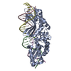
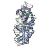
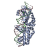
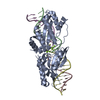
[English] 日本語
Yorodumi
- PDB-5e67: K103A/K262A double mutant of I-SmaMI -

+
Open data


ID or keywords:

Loading...

-
Basic information

Entry
Database: PDB / ID: 5.0E+67
TitleK103A/K262A double mutant of I-SmaMI
Components
  • DNA bottom strand
  • DNA top strand
  • I-SmaMI LAGLIDADG meganuclease
KeywordsHydrolase/DNA / LAGLIDADG / I-SmaMI / K103A/K262A / Hydrolase-DNA complex
Function / homology
Function and homology information


endonuclease activity / mitochondrion / metal ion binding
Similarity search - Function
LAGLIDADG endonuclease / Homing endonucleases / Endonuclease I-creI / Homing endonuclease, LAGLIDADG / Homing endonuclease / Roll / Alpha Beta
Similarity search - Domain/homology
2-METHOXYETHANOL / DNA / DNA (> 10) / Homing endonuclease LAGLIDADG domain-containing protein
Similarity search - Component
Biological speciesSordaria macrospora (fungus)
Synthetic construct (others)
MethodX-RAY DIFFRACTION / Resolution: 2.2 Å
AuthorsShen, B.W. / Stoddard, B.
Funding support United States, 1items
OrganizationGrant numberCountry
National Institutes of Health/National Institute of General Medical Sciences (NIH/NIGMS)RO1 GM United States
CitationJournal: J.Mol.Biol. / Year: 2016
Title: The Structural Basis of Asymmetry in DNA Binding and Cleavage as Exhibited by the I-SmaMI LAGLIDADG Meganuclease.
Authors: Shen, B.W. / Lambert, A. / Walker, B.C. / Stoddard, B.L. / Kaiser, B.K.
History
DepositionOct 9, 2015Deposition site: RCSB / Processing site: RCSB
Revision 1.0Jan 13, 2016Provider: repository / Type: Initial release
Revision 1.1Feb 10, 2016Group: Database references
Revision 1.2Sep 13, 2017Group: Author supporting evidence / Database references / Derived calculations
Category: citation / pdbx_audit_support / pdbx_struct_oper_list
Item: _citation.journal_id_CSD / _pdbx_audit_support.funding_organization / _pdbx_struct_oper_list.symmetry_operation
Revision 1.3Dec 25, 2019Group: Author supporting evidence / Category: pdbx_audit_support / Item: _pdbx_audit_support.funding_organization
Revision 1.4Mar 6, 2024Group: Data collection / Database references / Derived calculations
Category: chem_comp_atom / chem_comp_bond ...chem_comp_atom / chem_comp_bond / database_2 / pdbx_struct_conn_angle / struct_conn
Item: _database_2.pdbx_DOI / _database_2.pdbx_database_accession ..._database_2.pdbx_DOI / _database_2.pdbx_database_accession / _pdbx_struct_conn_angle.ptnr1_auth_asym_id / _pdbx_struct_conn_angle.ptnr1_auth_comp_id / _pdbx_struct_conn_angle.ptnr1_auth_seq_id / _pdbx_struct_conn_angle.ptnr1_label_asym_id / _pdbx_struct_conn_angle.ptnr1_label_atom_id / _pdbx_struct_conn_angle.ptnr1_label_comp_id / _pdbx_struct_conn_angle.ptnr1_label_seq_id / _pdbx_struct_conn_angle.ptnr3_auth_asym_id / _pdbx_struct_conn_angle.ptnr3_auth_comp_id / _pdbx_struct_conn_angle.ptnr3_auth_seq_id / _pdbx_struct_conn_angle.ptnr3_label_asym_id / _pdbx_struct_conn_angle.ptnr3_label_atom_id / _pdbx_struct_conn_angle.ptnr3_label_comp_id / _pdbx_struct_conn_angle.ptnr3_label_seq_id / _pdbx_struct_conn_angle.value / _struct_conn.pdbx_dist_value / _struct_conn.ptnr1_auth_asym_id / _struct_conn.ptnr1_auth_comp_id / _struct_conn.ptnr1_auth_seq_id / _struct_conn.ptnr1_label_asym_id / _struct_conn.ptnr1_label_atom_id / _struct_conn.ptnr1_label_comp_id / _struct_conn.ptnr1_label_seq_id / _struct_conn.ptnr2_auth_asym_id / _struct_conn.ptnr2_auth_comp_id / _struct_conn.ptnr2_auth_seq_id / _struct_conn.ptnr2_label_asym_id / _struct_conn.ptnr2_label_atom_id / _struct_conn.ptnr2_label_comp_id / _struct_conn.ptnr2_label_seq_id

-
Structure visualization

Structure viewerMolecule:
MolmilJmol/JSmol

Downloads & links

-
Assembly

Deposited unit
A: I-SmaMI LAGLIDADG meganuclease
B: DNA bottom strand
C: DNA top strand
hetero molecules


Theoretical massNumber of molelcules
Total (without water)49,6387
Polymers49,3933
Non-polymers2454
Water3,387188
1


  • Idetical with deposited unit
  • defined by author&software
TypeNameSymmetry operationNumber
identity operation1_555x,y,z1
Buried area8490 Å2
ΔGint-57 kcal/mol
Surface area18790 Å2
MethodPISA
Unit cell
Length a, b, c (Å)60.518, 67.326, 98.572
Angle α, β, γ (deg.)90.00, 90.00, 90.00
Int Tables number19
Space group name H-MP212121

-
Components

-
Protein , 1 types, 1 molecules A

#1: Protein I-SmaMI LAGLIDADG meganuclease


Mass: 34036.438 Da / Num. of mol.: 1 / Fragment: UNP residues 114-415 / Mutation: K103A, L165N, K262A, M268Q
Source method: isolated from a genetically manipulated source
Source: (gene. exp.) Sordaria macrospora (strain ATCC MYA-333 / DSM 997 / K(L3346) / K-hell) (fungus)
Strain: ATCC MYA-333 / DSM 997 / K(L3346) / K-hell / Gene: SMAC_12671 / Plasmid: pET24d(+) / Production host: ESCHERICHIA COLI (E. coli) / Strain (production host): BL21(DE3)RIL+ / References: UniProt: F7WD42

-
DNA chain , 2 types, 2 molecules BC

#2: DNA chain DNA bottom strand


Mass: 7691.988 Da / Num. of mol.: 1 / Source method: obtained synthetically / Source: (synth.) Synthetic construct (others)
#3: DNA chain DNA top strand


Mass: 7664.945 Da / Num. of mol.: 1 / Source method: obtained synthetically / Source: (synth.) Synthetic construct (others)

-
Non-polymers , 4 types, 192 molecules

#4: Chemical ChemComp-MG / MAGNESIUM ION


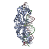
Mass: 24.305 Da / Num. of mol.: 2 / Source method: obtained synthetically / Formula: Mg
#5: Chemical ChemComp-MXE / 2-METHOXYETHANOL


Mass: 76.094 Da / Num. of mol.: 1 / Source method: obtained synthetically / Formula: C3H8O2
#6: Chemical ChemComp-PG0 / 2-(2-METHOXYETHOXY)ETHANOL / PEG 6000


Mass: 120.147 Da / Num. of mol.: 1 / Source method: obtained synthetically / Formula: C5H12O3 / Comment: inhibitor, precipitant*YM
#7: Water ChemComp-HOH / water


Mass: 18.015 Da / Num. of mol.: 188 / Source method: isolated from a natural source / Formula: H2O

-
Details

Sequence detailsMutation of L165N and M267Q were made to improve expression/purification.

-
Experimental details

-
Experiment

ExperimentMethod: X-RAY DIFFRACTION

-
Sample preparation

CrystalDensity Matthews: 2.22 Å3/Da / Density % sol: 39.49 %
Crystal growTemperature: 298 K / Method: vapor diffusion, hanging drop / pH: 7.5 / Details: 28 - 32% PEGMME550, HEPES, Mg++ / PH range: 7.5 - 8.0

-
Data collection

DiffractionMean temperature: 100 K
Diffraction sourceSource: ROTATING ANODE / Type: RIGAKU MICROMAX-007 HF / Wavelength: 1.54 Å
DetectorType: RIGAKU SATURN 70 / Detector: CCD / Date: Apr 15, 2015
RadiationProtocol: SINGLE WAVELENGTH / Monochromatic (M) / Laue (L): M / Scattering type: x-ray
Radiation wavelengthWavelength: 1.54 Å / Relative weight: 1
ReflectionResolution: 2.2→25 Å / Num. obs: 19836 / % possible obs: 92 % / Observed criterion σ(F): 1 / Observed criterion σ(I): 1 / Redundancy: 6.7 % / Rsym value: 0.088 / Net I/σ(I): 17
Reflection shellResolution: 2.2→2.28 Å / Redundancy: 4.4 % / Rmerge(I) obs: 0.619 / Mean I/σ(I) obs: 2.3 / % possible all: 59.2

-
Processing

Software
NameVersionClassification
REFMAC5.8.0124refinement
HKL-2000data reduction
SCALEPACKdata scaling
PHASERphasing
RefinementResolution: 2.2→25 Å / Cor.coef. Fo:Fc: 0.961 / Cor.coef. Fo:Fc free: 0.913 / SU B: 15.168 / SU ML: 0.189 / Cross valid method: THROUGHOUT / ESU R: 0.376 / ESU R Free: 0.256 / Stereochemistry target values: MAXIMUM LIKELIHOOD / Details: HYDROGENS HAVE BEEN ADDED IN THE RIDING POSITIONS
RfactorNum. reflection% reflectionSelection details
Rfree0.25448 957 4.9 %RANDOM
Rwork0.17909 ---
obs0.18276 18593 92.73 %-
Solvent computationIon probe radii: 0.8 Å / Shrinkage radii: 0.8 Å / VDW probe radii: 1.2 Å / Solvent model: MASK
Displacement parametersBiso mean: 34.571 Å2
Baniso -1Baniso -2Baniso -3
1-0.68 Å20 Å20 Å2
2---0.66 Å20 Å2
3----0.02 Å2
Refinement stepCycle: 1 / Resolution: 2.2→25 Å
ProteinNucleic acidLigandSolventTotal
Num. atoms2339 1019 15 188 3561
Refine LS restraints
Refine-IDTypeDev idealDev ideal targetNumber
X-RAY DIFFRACTIONr_bond_refined_d0.0180.0173563
X-RAY DIFFRACTIONr_bond_other_d0.0020.022958
X-RAY DIFFRACTIONr_angle_refined_deg2.0641.6925011
X-RAY DIFFRACTIONr_angle_other_deg1.30436869
X-RAY DIFFRACTIONr_dihedral_angle_1_deg6.9915295
X-RAY DIFFRACTIONr_dihedral_angle_2_deg37.32224.38898
X-RAY DIFFRACTIONr_dihedral_angle_3_deg17.60115449
X-RAY DIFFRACTIONr_dihedral_angle_4_deg17.627158
X-RAY DIFFRACTIONr_chiral_restr0.1170.2505
X-RAY DIFFRACTIONr_gen_planes_refined0.0110.023255
X-RAY DIFFRACTIONr_gen_planes_other0.0020.02792
X-RAY DIFFRACTIONr_nbd_refined
X-RAY DIFFRACTIONr_nbd_other
X-RAY DIFFRACTIONr_nbtor_refined
X-RAY DIFFRACTIONr_nbtor_other
X-RAY DIFFRACTIONr_xyhbond_nbd_refined
X-RAY DIFFRACTIONr_xyhbond_nbd_other
X-RAY DIFFRACTIONr_metal_ion_refined
X-RAY DIFFRACTIONr_metal_ion_other
X-RAY DIFFRACTIONr_symmetry_vdw_refined
X-RAY DIFFRACTIONr_symmetry_vdw_other
X-RAY DIFFRACTIONr_symmetry_hbond_refined
X-RAY DIFFRACTIONr_symmetry_hbond_other
X-RAY DIFFRACTIONr_symmetry_metal_ion_refined
X-RAY DIFFRACTIONr_symmetry_metal_ion_other
X-RAY DIFFRACTIONr_mcbond_it1.3792.0341180
X-RAY DIFFRACTIONr_mcbond_other1.382.0311179
X-RAY DIFFRACTIONr_mcangle_it2.3033.0481475
X-RAY DIFFRACTIONr_mcangle_other2.3023.0511476
X-RAY DIFFRACTIONr_scbond_it1.5062.2192383
X-RAY DIFFRACTIONr_scbond_other1.5032.2192383
X-RAY DIFFRACTIONr_scangle_it
X-RAY DIFFRACTIONr_scangle_other2.463.2713537
X-RAY DIFFRACTIONr_long_range_B_refined4.6718.3134422
X-RAY DIFFRACTIONr_long_range_B_other4.6718.3214423
X-RAY DIFFRACTIONr_rigid_bond_restr
X-RAY DIFFRACTIONr_sphericity_free
X-RAY DIFFRACTIONr_sphericity_bonded
LS refinement shellResolution: 2.2→2.257 Å / Total num. of bins used: 20
RfactorNum. reflection% reflection
Rfree0.307 45 -
Rwork0.233 861 -
obs--59.64 %
Refinement TLS params.

Method: refined / Refine-ID: X-RAY DIFFRACTION

IDL112)L122)L132)L222)L232)L332)S11 (Å °)S12 (Å °)S13 (Å °)S21 (Å °)S22 (Å °)S23 (Å °)S31 (Å °)S32 (Å °)S33 (Å °)T112)T122)T132)T222)T232)T332)Origin x (Å)Origin y (Å)Origin z (Å)
10.47830.37850.18752.48691.50051.7188-0.0292-0.01630.0686-0.0770.01730.1646-0.1456-0.07660.01190.02120.01520.00150.05110.02030.02935.8802-1.16-0.9494
20.3319-0.199-0.48462.81472.89813.80310.0882-0.11530.07360.0688-0.0620.117-0.0931-0.2736-0.02620.15490.0391-0.02450.2252-0.01410.1613-4.2619-0.8369-0.6502
30.09710.32170.58951.74892.51134.0991-0.0009-0.0664-0.0088-0.2351-0.11990.2436-0.2977-0.35790.12080.23170.0242-0.01540.2246-0.0320.2245-4.3520.3497-0.8656
Refinement TLS group
IDRefine-IDRefine TLS-IDAuth asym-IDAuth seq-ID
1X-RAY DIFFRACTION1A7 - 300
2X-RAY DIFFRACTION2B1 - 25
3X-RAY DIFFRACTION3C1 - 25

+
About Yorodumi

-
News

-
Feb 9, 2022. New format data for meta-information of EMDB entries

New format data for meta-information of EMDB entries

  • Version 3 of the EMDB header file is now the official format.
  • The previous official version 1.9 will be removed from the archive.

Related info.:EMDB header

External links:wwPDB to switch to version 3 of the EMDB data model

-
Aug 12, 2020. Covid-19 info

Covid-19 info

URL: https://pdbjlvh1.pdbj.org/emnavi/covid19.php

New page: Covid-19 featured information page in EM Navigator.

Related info.:Covid-19 info / Mar 5, 2020. Novel coronavirus structure data

+
Mar 5, 2020. Novel coronavirus structure data

Novel coronavirus structure data

Related info.:Yorodumi Speices / Aug 12, 2020. Covid-19 info

External links:COVID-19 featured content - PDBj / Molecule of the Month (242):Coronavirus Proteases

+
Jan 31, 2019. EMDB accession codes are about to change! (news from PDBe EMDB page)

EMDB accession codes are about to change! (news from PDBe EMDB page)

  • The allocation of 4 digits for EMDB accession codes will soon come to an end. Whilst these codes will remain in use, new EMDB accession codes will include an additional digit and will expand incrementally as the available range of codes is exhausted. The current 4-digit format prefixed with “EMD-” (i.e. EMD-XXXX) will advance to a 5-digit format (i.e. EMD-XXXXX), and so on. It is currently estimated that the 4-digit codes will be depleted around Spring 2019, at which point the 5-digit format will come into force.
  • The EM Navigator/Yorodumi systems omit the EMD- prefix.

Related info.:Q: What is EMD? / ID/Accession-code notation in Yorodumi/EM Navigator

External links:EMDB Accession Codes are Changing Soon! / Contact to PDBj

+
Jul 12, 2017. Major update of PDB

Major update of PDB

  • wwPDB released updated PDB data conforming to the new PDBx/mmCIF dictionary.
  • This is a major update changing the version number from 4 to 5, and with Remediation, in which all the entries are updated.
  • In this update, many items about electron microscopy experimental information are reorganized (e.g. em_software).
  • Now, EM Navigator and Yorodumi are based on the updated data.

External links:wwPDB Remediation / Enriched Model Files Conforming to OneDep Data Standards Now Available in the PDB FTP Archive

-
Yorodumi

Thousand views of thousand structures

  • Yorodumi is a browser for structure data from EMDB, PDB, SASBDB, etc.
  • This page is also the successor to EM Navigator detail page, and also detail information page/front-end page for Omokage search.
  • The word "yorodu" (or yorozu) is an old Japanese word meaning "ten thousand". "mi" (miru) is to see.

Related info.:EMDB / PDB / SASBDB / Comparison of 3 databanks / Yorodumi Search / Aug 31, 2016. New EM Navigator & Yorodumi / Yorodumi Papers / Jmol/JSmol / Function and homology information / Changes in new EM Navigator and Yorodumi

Read more