 Movie
Movie Controller

[English] 日本語
 Yorodumi
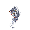
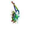
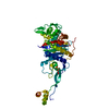
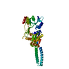
- PDB-3eqv: Crystal structure of penicillin-binding protein 2 from Neisseria ... -
+ Open data

- Basic information
| Entry | Database: PDB / ID: 3eqv | ||||||
|---|---|---|---|---|---|---|---|
| Title | Crystal structure of penicillin-binding protein 2 from Neisseria gonorrhoeae containing four mutations associated with penicillin resistance | ||||||
 Components Components | Penicillin-binding protein 2 | ||||||
 Keywords Keywords | BIOSYNTHETIC PROTEIN / Penicillin-binding protein / Class B transpeptidase / penicillin resistance / Cell division / Cell inner membrane / Cell membrane / Cell shape / Cell wall biogenesis/degradation / Peptidoglycan synthesis | ||||||
| Function / homology |  Function and homology information Function and homology informationpeptidoglycan glycosyltransferase activity / serine-type D-Ala-D-Ala carboxypeptidase / serine-type D-Ala-D-Ala carboxypeptidase activity / division septum assembly / FtsZ-dependent cytokinesis / penicillin binding / peptidoglycan biosynthetic process / cell wall organization / regulation of cell shape / response to antibiotic ...peptidoglycan glycosyltransferase activity / serine-type D-Ala-D-Ala carboxypeptidase / serine-type D-Ala-D-Ala carboxypeptidase activity / division septum assembly / FtsZ-dependent cytokinesis / penicillin binding / peptidoglycan biosynthetic process / cell wall organization / regulation of cell shape / response to antibiotic / proteolysis / plasma membrane Similarity search - Function | ||||||
| Biological species |  Neisseria gonorrhoeae (bacteria) Neisseria gonorrhoeae (bacteria) | ||||||
| Method |  X-RAY DIFFRACTION / SYNCHROTRON / FOURIER SYNTHESIS / Resolution: 2.4 Å X-RAY DIFFRACTION / SYNCHROTRON / FOURIER SYNTHESIS / Resolution: 2.4 Å | ||||||
 Authors Authors | Powell, A.J. / Deacon, A.M. / Nicholas, R.A. / Davies, C. | ||||||
 Citation Citation | Journal: J.Biol.Chem. / Year: 2009 Title: Crystal Structures of Penicillin-binding Protein 2 from Penicillin-susceptible and -resistant Strains of Neisseria gonorrhoeae Reveal an Unexpectedly Subtle Mechanism for Antibiotic Resistance. Authors: Powell, A.J. / Tomberg, J. / Deacon, A.M. / Nicholas, R.A. / Davies, C. | ||||||
| History |
|
- Structure visualization
| Structure viewer | Molecule: MolmilJmol/JSmol |
|---|
- Downloads & links
-Download
| PDBx/mmCIF format | 3eqv.cif.gz | 179.9 KB | Display | PDBx/mmCIF format |
|---|---|---|---|---|
| PDB format | pdb3eqv.ent.gz | 140.3 KB | Display | PDB format |
| PDBx/mmJSON format | 3eqv.json.gz | Tree view | PDBx/mmJSON format | |
| Others |  Other downloads Other downloads |
-Validation report
| Arichive directory | https://data.pdbj.org/pub/pdb/validation_reports/eq/3eqvftp://data.pdbj.org/pub/pdb/validation_reports/eq/3eqv | HTTPS FTP |
|---|
-Related structure data


-Links
 PDBj
PDBj
- Assembly
| Deposited unit | 
| ||||||||
|---|---|---|---|---|---|---|---|---|---|
| 1 | 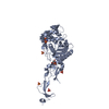
| ||||||||
| 2 | 
| ||||||||
| 3 | 
| ||||||||
| 4 | 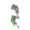
| ||||||||
| 5 |
| ||||||||
| Unit cell |
|
-Components
| #1: Protein | Mass: 59108.957 Da / Num. of mol.: 2 / Mutation: F504L, A510V, A516G, P551S Source method: isolated from a genetically manipulated source Source: (gene. exp.) Neisseria gonorrhoeae (bacteria) / Strain: 6140 / Gene: penA / Plasmid: pMAL-C2KV/H6 / Production host: References: UniProt: P08149, serine-type D-Ala-D-Ala carboxypeptidase #2: Chemical | ChemComp-SO4 /   Mass: 96.063 Da / Num. of mol.: 15 / Source method: obtained synthetically / Formula: SO4 Mass: 96.063 Da / Num. of mol.: 15 / Source method: obtained synthetically / Formula: SO4#3: Chemical |   Mass: 92.094 Da / Num. of mol.: 2 / Source method: obtained synthetically / Formula: C3H8O3 Mass: 92.094 Da / Num. of mol.: 2 / Source method: obtained synthetically / Formula: C3H8O3#4: Water | ChemComp-HOH / |  Mass: 18.015 Da / Num. of mol.: 79 / Source method: isolated from a natural source / Formula: H2O Mass: 18.015 Da / Num. of mol.: 79 / Source method: isolated from a natural source / Formula: H2OHas protein modification | Y | |
|---|
-Experimental details
-Experiment
| Experiment | Method: X-RAY DIFFRACTION / Number of used crystals: 1 |
|---|
- Sample preparation
| Crystal | Density Matthews: 3.84 Å3/Da / Density % sol: 67.97 % |
|---|---|
| Crystal grow | Temperature: 294 K / Method: vapor diffusion, hanging drop / pH: 8.5 Details: 2.2 M ammonium sulphate, 100 mM Tris-HCl, pH 8.5, VAPOR DIFFUSION, HANGING DROP, temperature 294K |
-Data collection
| Diffraction | Mean temperature: 100 K |
|---|---|
| Diffraction source | Source: SYNCHROTRON / Site: APS  / Beamline: 22-ID / Wavelength: 0.97934 Å / Beamline: 22-ID / Wavelength: 0.97934 Å |
| Detector | Type: MAR scanner 300 mm plate / Detector: IMAGE PLATE / Date: Nov 11, 2006 |
| Radiation | Monochromator: Si(220) / Protocol: SINGLE WAVELENGTH / Monochromatic (M) / Laue (L): M / Scattering type: x-ray |
| Radiation wavelength | Wavelength: 0.97934 Å / Relative weight: 1 |
| Reflection | Resolution: 2.4→50 Å / Num. all: 71399 / Num. obs: 71399 / % possible obs: 98 % / Observed criterion σ(F): 0 / Observed criterion σ(I): 0 / Redundancy: 7.1 % / Biso Wilson estimate: 49.1 Å2 / Rsym value: 0.101 / Net I/σ(I): 29.9 |
| Reflection shell | Resolution: 2.4→2.49 Å / Redundancy: 4.3 % / Mean I/σ(I) obs: 2.7 / Num. unique all: 5957 / Rsym value: 0.52 / % possible all: 83.5 |
- Processing
| Software |
| ||||||||||||||||||||||||||||||||||||||||||||||||||||||||||||||||||||||||||||||||||||||||||
|---|---|---|---|---|---|---|---|---|---|---|---|---|---|---|---|---|---|---|---|---|---|---|---|---|---|---|---|---|---|---|---|---|---|---|---|---|---|---|---|---|---|---|---|---|---|---|---|---|---|---|---|---|---|---|---|---|---|---|---|---|---|---|---|---|---|---|---|---|---|---|---|---|---|---|---|---|---|---|---|---|---|---|---|---|---|---|---|---|---|---|---|
| Refinement | Method to determine structure: FOURIER SYNTHESIS Starting model: Wild-type structure Resolution: 2.4→48.1 Å / Cor.coef. Fo:Fc: 0.938 / Cor.coef. Fo:Fc free: 0.921 / SU B: 15.651 / SU ML: 0.171 / Isotropic thermal model: isotropic / Cross valid method: THROUGHOUT / σ(F): 0 / ESU R: 0.219 / ESU R Free: 0.197 / Stereochemistry target values: MAXIMUM LIKELIHOOD
| ||||||||||||||||||||||||||||||||||||||||||||||||||||||||||||||||||||||||||||||||||||||||||
| Solvent computation | Ion probe radii: 0.8 Å / Shrinkage radii: 0.8 Å / VDW probe radii: 1.2 Å / Solvent model: MASK | ||||||||||||||||||||||||||||||||||||||||||||||||||||||||||||||||||||||||||||||||||||||||||
| Displacement parameters | Biso mean: 48.7 Å2
| ||||||||||||||||||||||||||||||||||||||||||||||||||||||||||||||||||||||||||||||||||||||||||
| Refinement step | Cycle: LAST / Resolution: 2.4→48.1 Å
| ||||||||||||||||||||||||||||||||||||||||||||||||||||||||||||||||||||||||||||||||||||||||||
| Refine LS restraints |
| ||||||||||||||||||||||||||||||||||||||||||||||||||||||||||||||||||||||||||||||||||||||||||
| LS refinement shell | Resolution: 2.4→2.454 Å / Total num. of bins used: 20
|
