Mass: 18.015 Da / Num. of mol.: 39 / Source method: isolated from a natural source / Formula: H2O
Has protein modification
Y
-
Experimental details
-
Experiment
Experiment
Method: X-RAY DIFFRACTION / Number of used crystals: 1
-
Sample preparation
Crystal
Density Matthews: 3.85 Å3/Da / Density % sol: 68 %
Crystal grow
Temperature: 297 K / Method: vapor diffusion, hanging drop / pH: 8.25 Details: Grown over reservoirs containing 2.2M ammonium sulfate buffered with 100 mM Tris pH 8.25
Resolution: 2.4→44.11 Å / Cor.coef. Fo:Fc: 0.937 / Cor.coef. Fo:Fc free: 0.922 / SU B: 14.524 / SU ML: 0.155 / Cross valid method: THROUGHOUT / ESU R: 0.22 / ESU R Free: 0.19 / Details: HYDROGENS HAVE BEEN USED IF PRESENT IN THE INPUT
Rfactor
Num. reflection
% reflection
Selection details
Rfree
0.244
3632
5.1 %
RANDOM
Rwork
0.218
-
-
-
obs
0.219
71799
98.4 %
-
Solvent computation
Ion probe radii: 0.8 Å / Shrinkage radii: 0.8 Å / VDW probe radii: 1.2 Å
 Movie
Movie Controller

 Yorodumi
 Open data
 Basic information
 Components
 Keywords
 Function and homology information
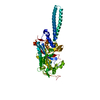
 Neisseria gonorrhoeae (bacteria)
 X-RAY DIFFRACTION /
  Authors
 United States, 1items
  Citation
 Structure visualization
 Downloads & links
 Other downloads

 PDBj
PDBj
 Assembly


 Mass: 92.094 Da / Num. of mol.: 3 / Source method: obtained synthetically / Formula: C3H8O3
 Mass: 96.063 Da / Num. of mol.: 12 / Source method: obtained synthetically / Formula: SO4
 Mass: 18.015 Da / Num. of mol.: 39 / Source method: isolated from a natural source / Formula: H2O
 Sample preparation
 Processing