 Movie
Movie Controller

[English] 日本語
 Yorodumi
- PDB-1p8k: The structure and DNA recognition of a bifunctional homing endonu... -
+ Open data

- Basic information
| Entry | Database: PDB / ID: 1p8k | ||||||
|---|---|---|---|---|---|---|---|
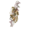
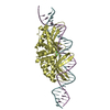
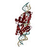
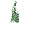
| Title | The structure and DNA recognition of a bifunctional homing endonuclease and group I intron splicing factor | ||||||
 Components Components |
| ||||||
 Keywords Keywords | HYDROLASE/DNA / HYDROLASE-DNA complex | ||||||
| Function / homology |  Function and homology information Function and homology informationintron homing / RNA splicing / mRNA processing / endonuclease activity / Hydrolases; Acting on ester bonds / hydrolase activity / mitochondrion Similarity search - Function | ||||||
| Biological species |  | ||||||
| Method |  X-RAY DIFFRACTION / SYNCHROTRON / SAD / Resolution: 2.6 Å X-RAY DIFFRACTION / SYNCHROTRON / SAD / Resolution: 2.6 Å | ||||||
 Authors Authors | Stoddard, B.L. / Bolduc, J.M. | ||||||
 Citation Citation | Journal: Genes Dev. / Year: 2003 Title: Structural and biochemical analyses of DNA and RNA binding by a bifunctional homing endonuclease and group I intron splicing factor. Authors: Bolduc, J.M. / Spiegel, P.C. / Chatterjee, P. / Brady, K.L. / Downing, M.E. / Caprara, M.G. / Waring, R.B. / Stoddard, B.L. | ||||||
| History |
| ||||||
| Remark 999 | SEQUENCE The author claims that Arg61 is correct. |
- Structure visualization
| Structure viewer | Molecule: MolmilJmol/JSmol |
|---|
- Downloads & links
-Download
| PDBx/mmCIF format | 1p8k.cif.gz | 100.9 KB | Display | PDBx/mmCIF format |
|---|---|---|---|---|
| PDB format | pdb1p8k.ent.gz | 72 KB | Display | PDB format |
| PDBx/mmJSON format | 1p8k.json.gz | Tree view | PDBx/mmJSON format | |
| Others |  Other downloads Other downloads |
-Validation report
| Arichive directory | https://data.pdbj.org/pub/pdb/validation_reports/p8/1p8kftp://data.pdbj.org/pub/pdb/validation_reports/p8/1p8k | HTTPS FTP |
|---|
-Related structure data
| Similar structure data |
|---|


-Links
 PDBj
PDBj


- Assembly
| Deposited unit | 
| ||||||||
|---|---|---|---|---|---|---|---|---|---|
| 1 |
| ||||||||
| Unit cell |
|
-Components
-DNA chain , 4 types, 4 molecules ABCD
| #1: DNA chain | Mass: 5588.601 Da / Num. of mol.: 1 / Source method: obtained synthetically |
|---|---|
| #2: DNA chain | Mass: 3975.611 Da / Num. of mol.: 1 / Source method: obtained synthetically |
| #3: DNA chain | Mass: 4931.230 Da / Num. of mol.: 1 / Source method: obtained synthetically |
| #4: DNA chain | Mass: 4481.899 Da / Num. of mol.: 1 / Source method: obtained synthetically |
-Protein / Non-polymers , 2 types, 3 molecules Z
| #5: Protein | Mass: 29615.154 Da / Num. of mol.: 1 Source method: isolated from a genetically manipulated source Source: (gene. exp.)  References: UniProt: P03880, Hydrolases; Acting on ester bonds |
|---|---|
| #6: Chemical |  Mass: 24.305 Da / Num. of mol.: 2 / Source method: obtained synthetically / Formula: Mg Mass: 24.305 Da / Num. of mol.: 2 / Source method: obtained synthetically / Formula: Mg |
-Details
| Has protein modification | Y |
|---|
-Experimental details
-Experiment
| Experiment | Method: X-RAY DIFFRACTION / Number of used crystals: 1 |
|---|
- Sample preparation
| Crystal | Density Matthews: 2.76 Å3/Da / Density % sol: 55.48 % | |||||||||||||||||||||||||||||||||||||||||||||||||
|---|---|---|---|---|---|---|---|---|---|---|---|---|---|---|---|---|---|---|---|---|---|---|---|---|---|---|---|---|---|---|---|---|---|---|---|---|---|---|---|---|---|---|---|---|---|---|---|---|---|---|
| Crystal grow | Temperature: 298 K / Method: vapor diffusion, hanging drop / pH: 5 Details: PEG3350, KCl, MgCl2, sodium citrate, VAPOR DIFFUSION, HANGING DROP, temperature 298K | |||||||||||||||||||||||||||||||||||||||||||||||||
| Components of the solutions |
| |||||||||||||||||||||||||||||||||||||||||||||||||
| Crystal grow | *PLUS Temperature: 22 ℃ / pH: 5 / Method: vapor diffusion | |||||||||||||||||||||||||||||||||||||||||||||||||
| Components of the solutions | *PLUS
|
-Data collection
| Diffraction | Mean temperature: 93 K |
|---|---|
| Diffraction source | Source: SYNCHROTRON / Site: ALS  / Beamline: 5.0.2 / Wavelength: 0.9794 Å / Beamline: 5.0.2 / Wavelength: 0.9794 Å |
| Detector | Type: ADSC QUANTUM 4 / Detector: CCD / Date: Nov 21, 2002 |
| Radiation | Monochromator: double crystal Si(111) / Protocol: SAD / Monochromatic (M) / Laue (L): M / Scattering type: x-ray |
| Radiation wavelength | Wavelength: 0.9794 Å / Relative weight: 1 |
| Reflection | Resolution: 2.6→50 Å / % possible obs: 90.4 % / Observed criterion σ(I): 0 / Redundancy: 3.5 % / Biso Wilson estimate: 40.1 Å2 / Rmerge(I) obs: 0.04 / Net I/σ(I): 9.5 |
| Reflection shell | Resolution: 2.6→2.69 Å / Rmerge(I) obs: 0.21 / % possible all: 61.8 |
| Reflection | *PLUS Num. obs: 16749 / Redundancy: 12.7 % / Num. measured all: 58621 / Rmerge(I) obs: 0.04 |
| Reflection shell | *PLUS % possible obs: 66.2 % / Rmerge(I) obs: 0.21 / Mean I/σ(I) obs: 3.2 |
- Processing
| Software |
| ||||||||||||||||||||
|---|---|---|---|---|---|---|---|---|---|---|---|---|---|---|---|---|---|---|---|---|---|
| Refinement | Method to determine structure: SAD / Resolution: 2.6→50 Å / σ(F): 0 / Stereochemistry target values: Engh & Huber
| ||||||||||||||||||||
| Refine analyze |
| ||||||||||||||||||||
| Refinement step | Cycle: LAST / Resolution: 2.6→50 Å
| ||||||||||||||||||||
| LS refinement shell | Resolution: 2.6→2.76 Å / Rfactor Rfree error: 0.03
| ||||||||||||||||||||
| Refinement | *PLUS Rfactor Rfree: 0.262 / Rfactor Rwork: 0.238 | ||||||||||||||||||||
| Solvent computation | *PLUS | ||||||||||||||||||||
| Displacement parameters | *PLUS | ||||||||||||||||||||
| Refine LS restraints | *PLUS
|
