- PDB-5dmk: Crystal Structure of IAg7 in complex with RLGL-WE14 -
+
Open data
ID or keywords:
Loading...
-
Basic information
Entry
Database: PDB / ID: 5dmk
Title
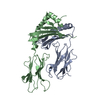
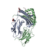
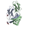
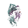
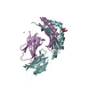
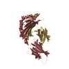
Crystal Structure of IAg7 in complex with RLGL-WE14
Components
H-2 class II histocompatibility antigen, A-D alpha chain
beta chain of Major Histocompatibility Complex Class II, I-Ag7,H2-Ab1 protein
Keywords
IMMUNE SYSTEM / Chromogranin A / Type I Diabetes / T cell / fusion protein
Function / homology
Function and homology information
protein localization to secretory granule / positive regulation of relaxation of cardiac muscle / Antimicrobial peptides / negative regulation of catecholamine secretion / positive regulation of dense core granule biogenesis / mast cell activation / mast cell chemotaxis / chromaffin granule / regulation of the force of heart contraction / positive regulation of T cell differentiation ...protein localization to secretory granule / positive regulation of relaxation of cardiac muscle / Antimicrobial peptides / negative regulation of catecholamine secretion / positive regulation of dense core granule biogenesis / mast cell activation / mast cell chemotaxis / chromaffin granule / regulation of the force of heart contraction / positive regulation of T cell differentiation / positive regulation of phospholipase C-activating G protein-coupled receptor signaling pathway / neuronal dense core vesicle / transport vesicle / multivesicular body / positive regulation of cardiac muscle contraction / secretory granule / mast cell degranulation / MHC class II protein complex / antigen processing and presentation of exogenous peptide antigen via MHC class II / peptide antigen binding / defense response to Gram-negative bacterium / adaptive immune response / early endosome / lysosome / defense response to Gram-positive bacterium / external side of plasma membrane / perinuclear region of cytoplasm / Golgi apparatus / cell surface / : Similarity search - Function
Chromogranin A/B / Chromogranin A/B/C / Chromogranin, conserved site / Granin (chromogranin or secretogranin) / Granins signature 1. / Granins signature 2. / Class II Histocompatibility Antigen, M Beta Chain; Chain B, domain 1 / Class II Histocompatibility Antigen, M Beta Chain; Chain B, domain 1 / MHC class II, beta chain, N-terminal / Class II histocompatibility antigen, beta domain ...Chromogranin A/B / Chromogranin A/B/C / Chromogranin, conserved site / Granin (chromogranin or secretogranin) / Granins signature 1. / Granins signature 2. / Class II Histocompatibility Antigen, M Beta Chain; Chain B, domain 1 / Class II Histocompatibility Antigen, M Beta Chain; Chain B, domain 1 / MHC class II, beta chain, N-terminal / Class II histocompatibility antigen, beta domain / Class II histocompatibility antigen, beta domain / MHC class II, alpha chain, N-terminal / Class II histocompatibility antigen, alpha domain / Class II histocompatibility antigen, alpha domain / MHC class II, alpha/beta chain, N-terminal / MHC classes I/II-like antigen recognition protein / : / Immunoglobulin/major histocompatibility complex, conserved site / Immunoglobulins and major histocompatibility complex proteins signature. / Immunoglobulin C-Type / Immunoglobulin C1-set / Immunoglobulin C1-set domain / Ig-like domain profile. / Immunoglobulin-like domain / Immunoglobulin-like domain superfamily / Roll / Immunoglobulin-like fold / Immunoglobulins / Immunoglobulin-like / Sandwich / Mainly Beta / Alpha Beta Similarity search - Domain/homology
CITRATE ANION / H-2 class II histocompatibility antigen, A-D alpha chain / Chromogranin-A / H2-Ab1 protein Similarity search - Component
Biological species
Mus musculus (house mouse)
Method
X-RAY DIFFRACTION / SYNCHROTRON / Resolution: 2.45 Å
#258 - Jun 2021 Glucocorticoid Receptor and Dexamethasone similarity (1)
#89 - May 2007 Aconitase and Iron Regulatory Protein 1 similarity (1)
-
Assembly
Deposited unit
A: H-2 class II histocompatibility antigen, A-D alpha chain B: beta chain of Major Histocompatibility Complex Class II, I-Ag7,H2-Ab1 protein C: H-2 class II histocompatibility antigen, A-D alpha chain D: beta chain of Major Histocompatibility Complex Class II, I-Ag7,H2-Ab1 protein E: H-2 class II histocompatibility antigen, A-D alpha chain F: beta chain of Major Histocompatibility Complex Class II, I-Ag7,H2-Ab1 protein G: H-2 class II histocompatibility antigen, A-D alpha chain H: beta chain of Major Histocompatibility Complex Class II, I-Ag7,H2-Ab1 protein hetero molecules
A: H-2 class II histocompatibility antigen, A-D alpha chain B: beta chain of Major Histocompatibility Complex Class II, I-Ag7,H2-Ab1 protein hetero molecules
E: H-2 class II histocompatibility antigen, A-D alpha chain F: beta chain of Major Histocompatibility Complex Class II, I-Ag7,H2-Ab1 protein hetero molecules
Mass: 18.015 Da / Num. of mol.: 333 / Source method: isolated from a natural source / Formula: H2O
Has protein modification
Y
Sequence details
Chain B,D,F,H are fusion protein, 19 residues of hybrid peptide (half from mouse Chromogranin A ...Chain B,D,F,H are fusion protein, 19 residues of hybrid peptide (half from mouse Chromogranin A gene, half from artificial peptide sequence library) linked to beta chain f of Chromogranin A
-
Experimental details
-
Experiment
Experiment
Method: X-RAY DIFFRACTION / Number of used crystals: 1
-
Sample preparation
Crystal
Density Matthews: 3.97 Å3/Da / Density % sol: 69.04 %
Crystal grow
Temperature: 298 K / Method: vapor diffusion, hanging drop / pH: 6.5 / Details: 1M Ammonium Citrate
In the structure databanks used in Yorodumi, some data are registered as the other names, "COVID-19 virus" and "2019-nCoV". Here are the details of the virus and the list of structure data.
Jan 31, 2019. EMDB accession codes are about to change! (news from PDBe EMDB page)
EMDB accession codes are about to change! (news from PDBe EMDB page)
The allocation of 4 digits for EMDB accession codes will soon come to an end. Whilst these codes will remain in use, new EMDB accession codes will include an additional digit and will expand incrementally as the available range of codes is exhausted. The current 4-digit format prefixed with “EMD-” (i.e. EMD-XXXX) will advance to a 5-digit format (i.e. EMD-XXXXX), and so on. It is currently estimated that the 4-digit codes will be depleted around Spring 2019, at which point the 5-digit format will come into force.
The EM Navigator/Yorodumi systems omit the EMD- prefix.
Related info.:Q: What is EMD? / ID/Accession-code notation in Yorodumi/EM Navigator
Yorodumi is a browser for structure data from EMDB, PDB, SASBDB, etc.
This page is also the successor to EM Navigator detail page, and also detail information page/front-end page for Omokage search.
The word "yorodu" (or yorozu) is an old Japanese word meaning "ten thousand". "mi" (miru) is to see.
Related info.:EMDB / PDB / SASBDB / Comparison of 3 databanks / Yorodumi Search / Aug 31, 2016. New EM Navigator & Yorodumi / Yorodumi Papers / Jmol/JSmol / Function and homology information / Changes in new EM Navigator and Yorodumi
 Movie
Movie Controller


 Open data
 Basic information
 Components
 Keywords
 Function and homology information
 X-RAY DIFFRACTION /
  Authors
 Citation
 Structure visualization
 Downloads & links
 Other downloads
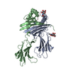


 PDBj
PDBj


 Assembly
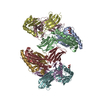


 Trichoplusia ni (cabbage looper) / References: UniProt: P04228
 Mass: 189.100 Da / Num. of mol.: 2 / Source method: obtained synthetically / Formula: C6H5O7
 Mass: 18.015 Da / Num. of mol.: 333 / Source method: isolated from a natural source / Formula: H2O
 Sample preparation
 / Beamline: 24-ID-E / Wavelength: 0.9795 Å
 Processing