 Movie
Movie Controller

[English] 日本語
 Yorodumi
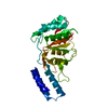
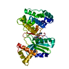
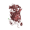
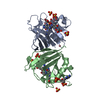
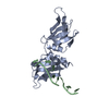
- PDB-5dmb: Crystal structure of a translational regulator bound to a flagell... -
+ Open data

- Basic information
| Entry | Database: PDB / ID: 5dmb | ||||||
|---|---|---|---|---|---|---|---|
| Title | Crystal structure of a translational regulator bound to a flagellar assembly factor | ||||||
 Components Components |
| ||||||
 Keywords Keywords | TRANSLATION / flagellum / assembly factor | ||||||
| Function / homology |  Function and homology information Function and homology informationregulation of bacterial-type flagellum assembly / regulation of carbohydrate metabolic process / bacterial-type flagellum organization / mRNA catabolic process / bacterial-type flagellum assembly / negative regulation of translational initiation / mRNA 5'-UTR binding / regulation of translation / cytoplasm / cytosol Similarity search - Function | ||||||
| Biological species |  Geobacillus thermodenitrificans (bacteria) Geobacillus thermodenitrificans (bacteria) | ||||||
| Method |  X-RAY DIFFRACTION / SYNCHROTRON / MOLECULAR REPLACEMENT / Resolution: 2.301 Å X-RAY DIFFRACTION / SYNCHROTRON / MOLECULAR REPLACEMENT / Resolution: 2.301 Å | ||||||
 Authors Authors | Altegoer, F. / Bange, G. | ||||||
 Citation Citation | Journal: Proc.Natl.Acad.Sci.USA / Year: 2016 Title: Structural basis for the CsrA-dependent modulation of translation initiation by an ancient regulatory protein. Authors: Altegoer, F. / Rensing, S.A. / Bange, G. | ||||||
| History |
|
- Structure visualization
| Structure viewer | Molecule: MolmilJmol/JSmol |
|---|
- Downloads & links
-Download
| PDBx/mmCIF format | 5dmb.cif.gz | 57.4 KB | Display | PDBx/mmCIF format |
|---|---|---|---|---|
| PDB format | pdb5dmb.ent.gz | 40.9 KB | Display | PDB format |
| PDBx/mmJSON format | 5dmb.json.gz | Tree view | PDBx/mmJSON format | |
| Others |  Other downloads Other downloads |
-Validation report
| Arichive directory | https://data.pdbj.org/pub/pdb/validation_reports/dm/5dmbftp://data.pdbj.org/pub/pdb/validation_reports/dm/5dmb | HTTPS FTP |
|---|
-Related structure data
| Related structure data | 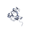 5dmdC 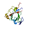 5jakC  2aj7S S: Starting model for refinement C: citing same article ( |
|---|---|
| Similar structure data |


-Links
 PDBj
PDBj- Assembly
| Deposited unit | 
| ||||||||
|---|---|---|---|---|---|---|---|---|---|
| 1 |
| ||||||||
| Unit cell |
|
-Components
| #1: Protein | Mass: 17389.801 Da / Num. of mol.: 1 Source method: isolated from a genetically manipulated source Source: (gene. exp.) Geobacillus thermodenitrificans (bacteria)Gene: fliW, GTNG_3059 / Plasmid: pET24d(+) / Production host: |
|---|---|
| #2: Protein | Mass: 9149.646 Da / Num. of mol.: 1 Source method: isolated from a genetically manipulated source Source: (gene. exp.) Geobacillus thermodenitrificans (bacteria)Gene: csrA, GTNG_3058 / Plasmid: pET16b / Production host: |
| #3: Water | ChemComp-HOH /  Mass: 18.015 Da / Num. of mol.: 73 / Source method: isolated from a natural source / Formula: H2O Mass: 18.015 Da / Num. of mol.: 73 / Source method: isolated from a natural source / Formula: H2O |
-Experimental details
-Experiment
| Experiment | Method: X-RAY DIFFRACTION |
|---|
- Sample preparation
| Crystal | Density Matthews: 2.81 Å3/Da / Density % sol: 56.27 % |
|---|---|
| Crystal grow | Temperature: 293 K / Method: vapor diffusion, sitting drop / Details: 0.2 M potassium fluoride, 20 % (w/v) PEG 3350 |
-Data collection
| Diffraction | Mean temperature: 100 K |
|---|---|
| Diffraction source | Source: SYNCHROTRON / Site: ESRF  / Beamline: MASSIF-1 / Wavelength: 0.966 Å / Beamline: MASSIF-1 / Wavelength: 0.966 Å |
| Detector | Type: PSI PILATUS 6M / Detector: PIXEL / Date: Dec 17, 2014 |
| Radiation | Protocol: SINGLE WAVELENGTH / Monochromatic (M) / Laue (L): M / Scattering type: x-ray |
| Radiation wavelength | Wavelength: 0.966 Å / Relative weight: 1 |
| Reflection | Resolution: 2.3→42.48 Å / Num. obs: 11552 / % possible obs: 99.5 % / Redundancy: 3.1 % / Rmerge(I) obs: 0.056 / Net I/σ(I): 1.8 |
| Reflection shell | Resolution: 2.3→2.42 Å / Rmerge(I) obs: 0.518 |
- Processing
| Software |
| |||||||||||||||||||||||||||||||||||
|---|---|---|---|---|---|---|---|---|---|---|---|---|---|---|---|---|---|---|---|---|---|---|---|---|---|---|---|---|---|---|---|---|---|---|---|---|
| Refinement | Method to determine structure: MOLECULAR REPLACEMENT Starting model: 2AJ7 Resolution: 2.301→42.477 Å / SU ML: 0.34 / Cross valid method: FREE R-VALUE / σ(F): 0 / Phase error: 37.18 / Stereochemistry target values: ML
| |||||||||||||||||||||||||||||||||||
| Solvent computation | Shrinkage radii: 0.9 Å / VDW probe radii: 1.11 Å / Solvent model: FLAT BULK SOLVENT MODEL | |||||||||||||||||||||||||||||||||||
| Refinement step | Cycle: LAST / Resolution: 2.301→42.477 Å
| |||||||||||||||||||||||||||||||||||
| Refine LS restraints |
| |||||||||||||||||||||||||||||||||||
| LS refinement shell |
|
