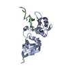
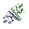
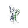
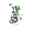
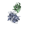
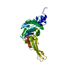
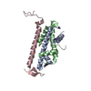
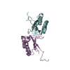
Entry Database : PDB / ID : 5ho4Title Crystal structure of hnRNPA2B1 in complex with 10-mer RNA Heterogeneous nuclear ribonucleoproteins A2/B1 RNA (5'-R(*AP*AP*GP*GP*AP*CP*UP*AP*GP*C)-3') Keywords / / / Function / homology Function Domain/homology Component
/ / / / / / / / / / / / / / / / / / / / / / / / / / / / / / / / / / / / / / / / / / / / / / / / / / / / / / / / / / / / / Biological species Homo sapiens (human)synthetic construct (others) Method / / Resolution : 1.85 Å Authors Wu, B.X. / Su, S.C. / Gan, J.H. / Ma, J.B. Journal : Nat Commun / Year : 2018Title : Molecular basis for the specific and multivariant recognitions of RNA substrates by human hnRNP A2/B1.Authors : Wu, B.X. / Su, S.C. / Patil, D.P. / Liu, H. / Gan, J.H. / Jaffrey, S.R. / Ma, J.B. History Deposition Jan 19, 2016 Deposition site / Processing site Revision 1.0 Feb 8, 2017 Provider / Type Revision 1.1 Mar 1, 2017 Group Revision 1.2 Feb 7, 2018 Group / Category / citation_authorItem _citation.country / _citation.journal_abbrev ... _citation.country / _citation.journal_abbrev / _citation.journal_id_CSD / _citation.journal_id_ISSN / _citation.journal_volume / _citation.pdbx_database_id_DOI / _citation.title / _citation.year Revision 1.3 May 22, 2019 Group / Database references / Structure summaryCategory / citation_author / structItem _citation.page_first / _citation.page_last ... _citation.page_first / _citation.page_last / _citation.pdbx_database_id_PubMed / _citation.title / _citation_author.identifier_ORCID / _struct.title Revision 1.4 Mar 20, 2024 Group / Database references / Category / chem_comp_bond / database_2Item / _database_2.pdbx_database_accession
Show all Show less
 Movie
Movie Controller


 Open data
 Basic information
 Components
 Keywords
 Function and homology information
 Homo sapiens (human)
 X-RAY DIFFRACTION /
  Authors
 Citation
 Structure visualization
 Downloads & links
 Other downloads
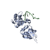


 PDBj
PDBj


 Assembly
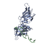
 Mass: 18.015 Da / Num. of mol.: 132 / Source method: isolated from a natural source / Formula: H2O
 Sample preparation
 / Beamline: BL18U1 / Wavelength: 0.987 Å
 Processing