 Movie
Movie Controller

[English] 日本語
 Yorodumi
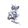
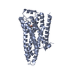
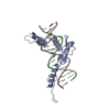
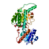
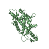
- PDB-1r7l: 2.0 A Crystal Structure of a Phage Protein from Bacillus cereus A... -
+ Open data

- Basic information
| Entry | Database: PDB / ID: 1r7l | ||||||
|---|---|---|---|---|---|---|---|
| Title | 2.0 A Crystal Structure of a Phage Protein from Bacillus cereus ATCC 14579 | ||||||
 Components Components | Phage protein | ||||||
 Keywords Keywords | STRUCTURAL GENOMICS / UNKNOWN FUNCTION / phage protein / two layers alpha-beta sandwich / PSI / Protein Structure Initiative / Midwest Center for Structural Genomics / MCSG | ||||||
| Function / homology | Bacillus phage protein / Bacillus phage protein-like / Bacillus phage protein-like / Phage ABA sandwich domain / Phage ABA sandwich domain / 2-Layer Sandwich / Alpha Beta / Phage protein Function and homology information Function and homology information | ||||||
| Biological species |  | ||||||
| Method |  X-RAY DIFFRACTION / SYNCHROTRON / MAD / Resolution: 2 Å X-RAY DIFFRACTION / SYNCHROTRON / MAD / Resolution: 2 Å | ||||||
 Authors Authors | Zhang, R. / Joachimiak, G. / Collart, F. / Joachimiak, A. / Midwest Center for Structural Genomics (MCSG) | ||||||
 Citation Citation | Journal: Proteins / Year: 2006 Title: Structure of phage protein BC1872 from Bacillus cereus, a singleton with new fold Authors: Zhang, R. / Joachimiak, G. / Jiang, S. / Cipriani, A. / Collart, F. / Joachimiak, A. | ||||||
| History |
|
- Structure visualization
| Structure viewer | Molecule: MolmilJmol/JSmol |
|---|
- Downloads & links
-Download
| PDBx/mmCIF format | 1r7l.cif.gz | 55.5 KB | Display | PDBx/mmCIF format |
|---|---|---|---|---|
| PDB format | pdb1r7l.ent.gz | 40.4 KB | Display | PDB format |
| PDBx/mmJSON format | 1r7l.json.gz | Tree view | PDBx/mmJSON format | |
| Others |  Other downloads Other downloads |
-Validation report
| Arichive directory | https://data.pdbj.org/pub/pdb/validation_reports/r7/1r7lftp://data.pdbj.org/pub/pdb/validation_reports/r7/1r7l | HTTPS FTP |
|---|
-Related structure data
| Similar structure data | |
|---|---|
| Other databases |


-Links
 PDBj
PDBj- Assembly
| Deposited unit | 
| ||||||||
|---|---|---|---|---|---|---|---|---|---|
| 1 |
| ||||||||
| Unit cell |
| ||||||||
| Details | Molecule A and molecule B represent the dimer in the asymmetric unit. |
-Components
| #1: Protein | Mass: 12498.479 Da / Num. of mol.: 2 Source method: isolated from a genetically manipulated source Source: (gene. exp.) #2: Water | ChemComp-HOH / |  Mass: 18.015 Da / Num. of mol.: 102 / Source method: isolated from a natural source / Formula: H2O Mass: 18.015 Da / Num. of mol.: 102 / Source method: isolated from a natural source / Formula: H2O |
|---|
-Experimental details
-Experiment
| Experiment | Method: X-RAY DIFFRACTION / Number of used crystals: 1 |
|---|
- Sample preparation
| Crystal | Density Matthews: 2.64 Å3/Da / Density % sol: 53.36 % |
|---|---|
| Crystal grow | Temperature: 298 K / Method: vapor diffusion, sitting drop / pH: 6 Details: 20% PEG 8000, 0.1M MES, 0.2M Ca(OAc)2, pH 6.0, VAPOR DIFFUSION, SITTING DROP, temperature 298K |
-Data collection
| Diffraction | Mean temperature: 100 K | ||||||||||||
|---|---|---|---|---|---|---|---|---|---|---|---|---|---|
| Diffraction source | Source: SYNCHROTRON / Site: APS  / Beamline: 19-ID / Wavelength: 0.9795, 0.9798, 0.9465 / Beamline: 19-ID / Wavelength: 0.9795, 0.9798, 0.9465 | ||||||||||||
| Detector | Type: SBC-2 / Detector: CCD / Date: Oct 20, 2003 / Details: mirrors | ||||||||||||
| Radiation | Monochromator: Si 111 channel / Protocol: MAD / Monochromatic (M) / Laue (L): M / Scattering type: x-ray | ||||||||||||
| Radiation wavelength |
| ||||||||||||
| Reflection | Resolution: 2→50 Å / Num. all: 17716 / Num. obs: 17557 / % possible obs: 99.1 % / Observed criterion σ(F): 4 / Observed criterion σ(I): 4 / Redundancy: 8.12 % / Biso Wilson estimate: 16.2 Å2 / Rmerge(I) obs: 0.098 / Net I/σ(I): 22.3 | ||||||||||||
| Reflection shell | Resolution: 2→2.07 Å / Redundancy: 7.2 % / Rmerge(I) obs: 0.518 / Mean I/σ(I) obs: 2.25 / % possible all: 95.6 |
- Processing
| Software |
| |||||||||||||||||||||||||
|---|---|---|---|---|---|---|---|---|---|---|---|---|---|---|---|---|---|---|---|---|---|---|---|---|---|---|
| Refinement | Method to determine structure: MAD / Resolution: 2→24.9 Å / Rfactor Rfree error: 0.007 / Data cutoff high absF: 444377.86 / Data cutoff low absF: 0 / Isotropic thermal model: RESTRAINED / Cross valid method: THROUGHOUT / σ(F): 0 / Stereochemistry target values: Engh & Huber / Details: Freidel pairs were used in the refinement.
| |||||||||||||||||||||||||
| Solvent computation | Solvent model: FLAT MODEL / Bsol: 60.0238 Å2 / ksol: 0.378568 e/Å3 | |||||||||||||||||||||||||
| Displacement parameters | Biso mean: 33.6 Å2
| |||||||||||||||||||||||||
| Refine analyze |
| |||||||||||||||||||||||||
| Refinement step | Cycle: LAST / Resolution: 2→24.9 Å
| |||||||||||||||||||||||||
| Refine LS restraints |
| |||||||||||||||||||||||||
| LS refinement shell | Resolution: 2→2.13 Å / Rfactor Rfree error: 0.023 / Total num. of bins used: 6
| |||||||||||||||||||||||||
| Xplor file |
|
