| Entry | Database: PDB / ID: 4lmo
|
|---|
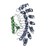
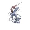
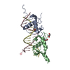
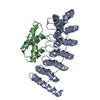
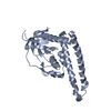
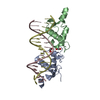
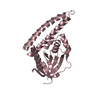
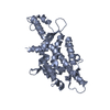
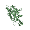
| Title | Structure of a vertebrate RNA binding domain of telomerase (TRBD) |
|---|
 Components Components | Telomerase reverse transcriptase |
|---|
 Keywords Keywords | RNA BINDING PROTEIN / RNA binding domain of the reverse transcriptase telomerase |
|---|
| Function / homology |  Function and homology information Function and homology information
DNA strand elongation / telomerase catalytic core complex / telomerase activity / telomerase RNA binding / telomeric repeat DNA binding / telomere maintenance via telomerase / RNA-directed DNA polymerase / chromosome, telomeric region / metal ion bindingSimilarity search - Function Topoisomerase I; Chain A, domain 4 - #70 / : / Telomerase reverse transcriptase, C-terminal extension / Telomerase ribonucleoprotein complex - RNA binding domain / Telomerase reverse transcriptase / Telomerase ribonucleoprotein complex - RNA-binding domain / Telomerase ribonucleoprotein complex - RNA binding domain / Topoisomerase I; Chain A, domain 4 / Reverse transcriptase (RNA-dependent DNA polymerase) / Reverse transcriptase domain ...Topoisomerase I; Chain A, domain 4 - #70 / : / Telomerase reverse transcriptase, C-terminal extension / Telomerase ribonucleoprotein complex - RNA binding domain / Telomerase reverse transcriptase / Telomerase ribonucleoprotein complex - RNA-binding domain / Telomerase ribonucleoprotein complex - RNA binding domain / Topoisomerase I; Chain A, domain 4 / Reverse transcriptase (RNA-dependent DNA polymerase) / Reverse transcriptase domain / Reverse transcriptase (RT) catalytic domain profile. / DNA/RNA polymerase superfamily / Orthogonal Bundle / Mainly AlphaSimilarity search - Domain/homology |
|---|
| Biological species |   Takifugu rubripes (torafugu) Takifugu rubripes (torafugu) |
|---|
| Method |  X-RAY DIFFRACTION / SYNCHROTRON / MAD / Resolution: 2.37 Å X-RAY DIFFRACTION / SYNCHROTRON / MAD / Resolution: 2.37 Å |
|---|
 Authors Authors | Harkisheimer, M. / Mason, M. / Shuvaeva, E. / Skordalakes, E. |
|---|
 Citation Citation | Journal: Structure / Year: 2013
Title: A Motif in the Vertebrate Telomerase N-Terminal Linker of TERT Contributes to RNA Binding and Telomerase Activity and Processivity.
Authors: Harkisheimer, M. / Mason, M. / Shuvaeva, E. / Skordalakes, E. |
|---|
| History | | Deposition | Jul 10, 2013 | Deposition site: RCSB / Processing site: RCSB |
|---|
| Revision 1.0 | Oct 9, 2013 | Provider: repository / Type: Initial release |
|---|
| Revision 1.1 | Oct 30, 2013 | Group: Database references |
|---|
| Revision 1.2 | Nov 20, 2024 | Group: Data collection / Database references / Structure summary
Category: chem_comp_atom / chem_comp_bond ...chem_comp_atom / chem_comp_bond / database_2 / pdbx_entry_details / pdbx_modification_feature / struct_ref_seq_dif
Item: _database_2.pdbx_DOI / _database_2.pdbx_database_accession / _struct_ref_seq_dif.details |
|---|
|
|---|
 Movie
Movie Controller

 Yorodumi
 Open data
 Basic information
 Structure visualization
 Downloads & links
 Other downloads


 PDBj
PDBj
 Assembly
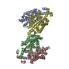


 Mass: 18.015 Da / Num. of mol.: 144 / Source method: isolated from a natural source / Formula: H2O
 Sample preparation
 Processing