[English] 日本語
Yorodumi
- PDB-3aji: Structure of Gankyrin-S6ATPase photo-cross-linked site-specifical... -

+
Open data


ID or keywords:

Loading...

-
Basic information

Entry
Database: PDB / ID: 3aji
TitleStructure of Gankyrin-S6ATPase photo-cross-linked site-specifically, and incoporated by genetic code expansion
Components
  • 26S proteasome non-ATPase regulatory subunit 10
  • Proteasome (Prosome, macropain) 26S subunit, ATPase, 4
KeywordsCHAPERONE/PROTEIN BINDING / Gankyrin / S6 atpase / p-benzoyl-L-phenylalanine / pbpa / amber suppression / Structural Genomics / RIKEN Structural Genomics/Proteomics Initiative / RSGI / CHAPERONE-PROTEIN BINDING complex
Function / homology
Function and homology information


Regulation of ornithine decarboxylase (ODC) / Proteasome assembly / Cross-presentation of soluble exogenous antigens (endosomes) / Autodegradation of Cdh1 by Cdh1:APC/C / APC/C:Cdc20 mediated degradation of Securin / Ubiquitin-Mediated Degradation of Phosphorylated Cdc25A / Ubiquitin-dependent degradation of Cyclin D / AUF1 (hnRNP D0) binds and destabilizes mRNA / Cdc20:Phospho-APC/C mediated degradation of Cyclin A / SCF(Skp2)-mediated degradation of p27/p21 ...Regulation of ornithine decarboxylase (ODC) / Proteasome assembly / Cross-presentation of soluble exogenous antigens (endosomes) / Autodegradation of Cdh1 by Cdh1:APC/C / APC/C:Cdc20 mediated degradation of Securin / Ubiquitin-Mediated Degradation of Phosphorylated Cdc25A / Ubiquitin-dependent degradation of Cyclin D / AUF1 (hnRNP D0) binds and destabilizes mRNA / Cdc20:Phospho-APC/C mediated degradation of Cyclin A / SCF(Skp2)-mediated degradation of p27/p21 / Assembly of the pre-replicative complex / CDK-mediated phosphorylation and removal of Cdc6 / Autodegradation of the E3 ubiquitin ligase COP1 / G2/M Checkpoints / Degradation of AXIN / APC/C:Cdh1 mediated degradation of Cdc20 and other APC/C:Cdh1 targeted proteins in late mitosis/early G1 / Asymmetric localization of PCP proteins / Regulation of RUNX3 expression and activity / Regulation of RAS by GAPs / Regulation of PTEN stability and activity / Regulation of RUNX2 expression and activity / Degradation of GLI1 by the proteasome / UCH proteinases / FBXL7 down-regulates AURKA during mitotic entry and in early mitosis / Degradation of DVL / Orc1 removal from chromatin / GSK3B and BTRC:CUL1-mediated-degradation of NFE2L2 / Dectin-1 mediated noncanonical NF-kB signaling / NIK-->noncanonical NF-kB signaling / TNFR2 non-canonical NF-kB pathway / Hedgehog ligand biogenesis / Hedgehog 'on' state / Oxygen-dependent proline hydroxylation of Hypoxia-inducible Factor Alpha / Degradation of beta-catenin by the destruction complex / Activation of NF-kappaB in B cells / The role of GTSE1 in G2/M progression after G2 checkpoint / FCERI mediated NF-kB activation / CLEC7A (Dectin-1) signaling / Interleukin-1 signaling / RUNX1 regulates transcription of genes involved in differentiation of HSCs / proteasome regulatory particle assembly / Downstream TCR signaling / Separation of Sister Chromatids / MAPK6/MAPK4 signaling / GLI3 is processed to GLI3R by the proteasome / ABC-family proteins mediated transport / Neddylation / proteasome accessory complex / positive regulation of cyclin-dependent protein serine/threonine kinase activity / Ub-specific processing proteases / KEAP1-NFE2L2 pathway / cytosolic proteasome complex / Antigen processing: Ubiquitination & Proteasome degradation / proteasome regulatory particle, base subcomplex / negative regulation of NF-kappaB transcription factor activity / negative regulation of release of cytochrome c from mitochondria / negative regulation of DNA damage response, signal transduction by p53 class mediator / blastocyst development / negative regulation of MAPK cascade / inclusion body / positive regulation of protein ubiquitination / positive regulation of proteasomal ubiquitin-dependent protein catabolic process / microtubule cytoskeleton / positive regulation of cell growth / ciliary basal body / cilium / apoptotic process / negative regulation of apoptotic process / negative regulation of transcription by RNA polymerase II / ATP hydrolysis activity / nucleoplasm / ATP binding / nucleus / cytosol
Similarity search - Function
: / : / Proteasomal ATPase OB C-terminal domain / Proteasomal ATPase OB C-terminal domain / Helicase, Ruva Protein; domain 3 - #60 / Ankyrin repeat-containing domain / AAA ATPase, AAA+ lid domain / AAA+ lid domain / ATPase, AAA-type, conserved site / AAA-protein family signature. ...: / : / Proteasomal ATPase OB C-terminal domain / Proteasomal ATPase OB C-terminal domain / Helicase, Ruva Protein; domain 3 - #60 / Ankyrin repeat-containing domain / AAA ATPase, AAA+ lid domain / AAA+ lid domain / ATPase, AAA-type, conserved site / AAA-protein family signature. / Helicase, Ruva Protein; domain 3 / Ankyrin repeat / Ankyrin repeats (3 copies) / ATPase family associated with various cellular activities (AAA) / Ankyrin repeat profile. / Ankyrin repeat region circular profile. / ankyrin repeats / ATPase, AAA-type, core / Ankyrin repeat / Ankyrin repeat-containing domain superfamily / Serine Threonine Protein Phosphatase 5, Tetratricopeptide repeat / Alpha Horseshoe / Nucleic acid-binding, OB-fold / ATPases associated with a variety of cellular activities / AAA+ ATPase domain / P-loop containing nucleoside triphosphate hydrolase / Orthogonal Bundle / Mainly Alpha
Similarity search - Domain/homology
26S proteasome regulatory subunit 6B / 26S proteasome regulatory subunit 6B / 26S proteasome non-ATPase regulatory subunit 10
Similarity search - Component
Biological speciesMus musculus (house mouse)
MethodX-RAY DIFFRACTION / SYNCHROTRON / MOLECULAR REPLACEMENT / Resolution: 2.05 Å
AuthorsSato, S. / Mimasu, S. / Sato, A. / Hino, N. / Sakamoto, K. / Umehara, T. / Yokoyama, S. / RIKEN Structural Genomics/Proteomics Initiative (RSGI)
CitationJournal: Biochemistry / Year: 2011
Title: Crystallographic study of a site-specifically cross-linked protein complex with a genetically incorporated photoreactive amino acid
Authors: Sato, S. / Mimasu, S. / Sato, A. / Hino, N. / Sakamoto, K. / Umehara, T. / Yokoyama, S.
History
DepositionJun 7, 2010Deposition site: PDBJ / Processing site: PDBJ
Revision 1.0Dec 22, 2010Provider: repository / Type: Initial release
Revision 1.1Jul 13, 2011Group: Version format compliance
Revision 1.2Jan 29, 2014Group: Database references
Revision 1.3Nov 1, 2023Group: Data collection / Database references ...Data collection / Database references / Derived calculations / Refinement description
Category: chem_comp_atom / chem_comp_bond ...chem_comp_atom / chem_comp_bond / database_2 / pdbx_initial_refinement_model / struct_conn / struct_ref_seq_dif
Item: _database_2.pdbx_DOI / _database_2.pdbx_database_accession ..._database_2.pdbx_DOI / _database_2.pdbx_database_accession / _struct_conn.pdbx_leaving_atom_flag / _struct_ref_seq_dif.details
Revision 1.4Nov 15, 2023Group: Data collection / Category: chem_comp_atom / chem_comp_bond / Item: _chem_comp_atom.atom_id / _chem_comp_bond.atom_id_2

-
Structure visualization

Structure viewerMolecule:
MolmilJmol/JSmol

Downloads & links

-
Assembly

Deposited unit
A: 26S proteasome non-ATPase regulatory subunit 10
B: Proteasome (Prosome, macropain) 26S subunit, ATPase, 4
C: 26S proteasome non-ATPase regulatory subunit 10
D: Proteasome (Prosome, macropain) 26S subunit, ATPase, 4


Theoretical massNumber of molelcules
Total (without water)69,9504
Polymers69,9504
Non-polymers00
Water5,891327
1
A: 26S proteasome non-ATPase regulatory subunit 10
B: Proteasome (Prosome, macropain) 26S subunit, ATPase, 4


Theoretical massNumber of molelcules
Total (without water)34,9752
Polymers34,9752
Non-polymers00
Water362
TypeNameSymmetry operationNumber
identity operation1_555x,y,z1
Buried area2340 Å2
ΔGint1 kcal/mol
Surface area13260 Å2
MethodPISA
2
C: 26S proteasome non-ATPase regulatory subunit 10
D: Proteasome (Prosome, macropain) 26S subunit, ATPase, 4


Theoretical massNumber of molelcules
Total (without water)34,9752
Polymers34,9752
Non-polymers00
Water362
TypeNameSymmetry operationNumber
identity operation1_555x,y,z1
Buried area2260 Å2
ΔGint0 kcal/mol
Surface area13390 Å2
MethodPISA
Unit cell
Length a, b, c (Å)103.164, 103.164, 154.993
Angle α, β, γ (deg.)90.00, 90.00, 120.00
Int Tables number146
Space group name H-MH3
Components on special symmetry positions
IDModelComponents
11A-253-

HOH

21C-263-

HOH

31C-266-

HOH

41D-238-

HOH

-
Components

#1: Protein 26S proteasome non-ATPase regulatory subunit 10 / 26S proteasome regulatory subunit p28 / Gankyrin


Mass: 25181.635 Da / Num. of mol.: 2 / Mutation: R85F
Source method: isolated from a genetically manipulated source
Source: (gene. exp.) Mus musculus (house mouse) / Gene: Psmd10 / Production host: Escherichia coli (E. coli) / References: UniProt: Q9Z2X2
#2: Protein Proteasome (Prosome, macropain) 26S subunit, ATPase, 4 / S6C


Mass: 9793.156 Da / Num. of mol.: 2 / Fragment: C-terminal domain
Source method: isolated from a genetically manipulated source
Source: (gene. exp.) Mus musculus (house mouse) / Gene: Psmc4 / Production host: Escherichia coli (E. coli) / References: UniProt: Q6ZWN9, UniProt: P54775*PLUS
#3: Water ChemComp-HOH / water


Mass: 18.015 Da / Num. of mol.: 327 / Source method: isolated from a natural source / Formula: H2O
Sequence details
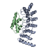
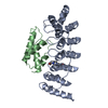
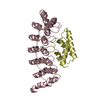
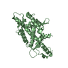
THE 85TH NON-NATURAL RESIDUE IS THE PHOTO REACTED VERSION OF PBPA INCOPORATED BY THE GENETIC CODE ...THE 85TH NON-NATURAL RESIDUE IS THE PHOTO REACTED VERSION OF PBPA INCOPORATED BY THE GENETIC CODE EXPANSION, AND THE 356TH RESIDUE IS THE PHOTO-COVALENT-BONDED PBPA ON GLUTAMIC ACID.

-
Experimental details

-
Experiment

ExperimentMethod: X-RAY DIFFRACTION / Number of used crystals: 2

-
Sample preparation

CrystalDensity Matthews: 2.27 Å3/Da / Density % sol: 45.8 %
Crystal growTemperature: 298 K / Method: vapor diffusion, hanging drop / pH: 8.5
Details: 31% polyethylene glycol 4000, 0.26M MgCl2, pH 8.5, VAPOR DIFFUSION, HANGING DROP, temperature 298.0K

-
Data collection

Diffraction
IDMean temperature (K)Crystal-ID
11001
21001
Diffraction source
SourceSiteBeamlineID
SYNCHROTRONSPring-8 BL41XU1
SYNCHROTRONSPring-8 BL26B22
Detector
TypeIDDetectorDate
RAYONIX MX225HE1CCDDec 15, 2008
RAYONIX MX-2252CCDOct 24, 2008
Radiation
IDProtocolMonochromatic (M) / Laue (L)Scattering typeWavelength-ID
1SINGLE WAVELENGTHMx-ray1
2SINGLE WAVELENGTHMx-ray1
Radiation wavelengthRelative weight: 1
ReflectionResolution: 2.05→50 Å / Num. obs: 38604 / % possible obs: 100 % / Redundancy: 4.2 % / Biso Wilson estimate: 26.5 Å2 / Rsym value: 0.098 / Net I/σ(I): 20.7
Reflection shellResolution: 2.05→2.18 Å / Redundancy: 4.2 % / Mean I/σ(I) obs: 3.2 / Num. unique all: 6083 / Rsym value: 0.454 / % possible all: 99.9

-
Processing

Software
NameVersionClassification
HKL-2000data collection
MOLREPphasing
CNS1.2refinement
DENZOdata reduction
SCALEPACKdata scaling
RefinementMethod to determine structure: MOLECULAR REPLACEMENT
Starting model: PDB ENTRY 2DVW
Resolution: 2.05→42.93 Å / Rfactor Rfree error: 0.005 / Data cutoff high absF: 2665091.25 / Data cutoff low absF: 0 / Isotropic thermal model: RESTRAINED / Cross valid method: THROUGHOUT / σ(F): 0 / σ(I): -3 / Details: BULK SOLVENT MODEL USED
RfactorNum. reflection% reflectionSelection details
Rfree0.229 1914 5 %RANDOM
Rwork0.171 ---
obs0.171 38604 100 %-
Solvent computationSolvent model: FLAT MODEL / Bsol: 62.4515 Å2 / ksol: 0.4 e/Å3
Displacement parametersBiso mean: 37.4 Å2
Baniso -1Baniso -2Baniso -3
1-4.13 Å20 Å20 Å2
2--4.13 Å20 Å2
3----8.27 Å2
Refine analyze
FreeObs
Luzzati coordinate error0.27 Å0.19 Å
Luzzati d res low-5 Å
Luzzati sigma a0.24 Å0.14 Å
Refinement stepCycle: LAST / Resolution: 2.05→42.93 Å
ProteinNucleic acidLigandSolventTotal
Num. atoms4668 0 0 327 4995
Refine LS restraints
Refine-IDTypeDev ideal
X-RAY DIFFRACTIONc_bond_d0.032
X-RAY DIFFRACTIONc_bond_d_na
X-RAY DIFFRACTIONc_bond_d_prot
X-RAY DIFFRACTIONc_angle_d
X-RAY DIFFRACTIONc_angle_d_na
X-RAY DIFFRACTIONc_angle_d_prot
X-RAY DIFFRACTIONc_angle_deg2.8
X-RAY DIFFRACTIONc_angle_deg_na
X-RAY DIFFRACTIONc_angle_deg_prot
X-RAY DIFFRACTIONc_dihedral_angle_d21.9
X-RAY DIFFRACTIONc_dihedral_angle_d_na
X-RAY DIFFRACTIONc_dihedral_angle_d_prot
X-RAY DIFFRACTIONc_improper_angle_d1.78
X-RAY DIFFRACTIONc_improper_angle_d_na
X-RAY DIFFRACTIONc_improper_angle_d_prot
X-RAY DIFFRACTIONc_mcbond_it
X-RAY DIFFRACTIONc_mcangle_it
X-RAY DIFFRACTIONc_scbond_it
X-RAY DIFFRACTIONc_scangle_it
LS refinement shellResolution: 2.05→2.18 Å / Rfactor Rfree error: 0.015 / Total num. of bins used: 6
RfactorNum. reflection% reflection
Rfree0.283 347 5.4 %
Rwork0.208 6083 -
obs-6083 99.9 %
Xplor file
Refine-IDSerial noParam fileTopol file
X-RAY DIFFRACTION1protein_rep.paramprotein.top
X-RAY DIFFRACTION2pbpa.parampbpa.top
X-RAY DIFFRACTION3water_rep.param

+
About Yorodumi

-
News

-
Feb 9, 2022. New format data for meta-information of EMDB entries

New format data for meta-information of EMDB entries

  • Version 3 of the EMDB header file is now the official format.
  • The previous official version 1.9 will be removed from the archive.

Related info.:EMDB header

External links:wwPDB to switch to version 3 of the EMDB data model

-
Aug 12, 2020. Covid-19 info

Covid-19 info

URL: https://pdbj.org/emnavi/covid19.php

New page: Covid-19 featured information page in EM Navigator.

Related info.:Covid-19 info / Mar 5, 2020. Novel coronavirus structure data

+
Mar 5, 2020. Novel coronavirus structure data

Novel coronavirus structure data

Related info.:Yorodumi Speices / Aug 12, 2020. Covid-19 info

External links:COVID-19 featured content - PDBj / Molecule of the Month (242):Coronavirus Proteases

+
Jan 31, 2019. EMDB accession codes are about to change! (news from PDBe EMDB page)

EMDB accession codes are about to change! (news from PDBe EMDB page)

  • The allocation of 4 digits for EMDB accession codes will soon come to an end. Whilst these codes will remain in use, new EMDB accession codes will include an additional digit and will expand incrementally as the available range of codes is exhausted. The current 4-digit format prefixed with “EMD-” (i.e. EMD-XXXX) will advance to a 5-digit format (i.e. EMD-XXXXX), and so on. It is currently estimated that the 4-digit codes will be depleted around Spring 2019, at which point the 5-digit format will come into force.
  • The EM Navigator/Yorodumi systems omit the EMD- prefix.

Related info.:Q: What is EMD? / ID/Accession-code notation in Yorodumi/EM Navigator

External links:EMDB Accession Codes are Changing Soon! / Contact to PDBj

+
Jul 12, 2017. Major update of PDB

Major update of PDB

  • wwPDB released updated PDB data conforming to the new PDBx/mmCIF dictionary.
  • This is a major update changing the version number from 4 to 5, and with Remediation, in which all the entries are updated.
  • In this update, many items about electron microscopy experimental information are reorganized (e.g. em_software).
  • Now, EM Navigator and Yorodumi are based on the updated data.

External links:wwPDB Remediation / Enriched Model Files Conforming to OneDep Data Standards Now Available in the PDB FTP Archive

-
Yorodumi

Thousand views of thousand structures

  • Yorodumi is a browser for structure data from EMDB, PDB, SASBDB, etc.
  • This page is also the successor to EM Navigator detail page, and also detail information page/front-end page for Omokage search.
  • The word "yorodu" (or yorozu) is an old Japanese word meaning "ten thousand". "mi" (miru) is to see.

Related info.:EMDB / PDB / SASBDB / Comparison of 3 databanks / Yorodumi Search / Aug 31, 2016. New EM Navigator & Yorodumi / Yorodumi Papers / Jmol/JSmol / Function and homology information / Changes in new EM Navigator and Yorodumi

Read more