- PDB-2dwz: Structure of the Oncoprotein Gankyrin in Complex with S6 ATPase o... -
+
Open data
ID or keywords:
Loading...
-
Basic information
Entry
Database: PDB / ID: 2dwz
Title
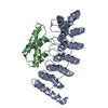
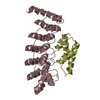
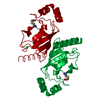
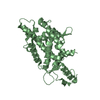
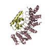
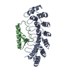
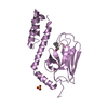
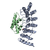
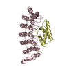
Structure of the Oncoprotein Gankyrin in Complex with S6 ATPase of the 26S Proteasome
Components
26S protease regulatory subunit 6B
26S proteasome non-ATPase regulatory subunit 10
Keywords
ONCOPROTEIN / ANKYRIN REPEATS / A-HELICAL DOMAIN / Structural Genomics / NPPSFA / National Project on Protein Structural and Functional Analyses / RIKEN Structural Genomics/Proteomics Initiative / RSGI
Function / homology
Function and homology information
Cross-presentation of soluble exogenous antigens (endosomes) / Proteasome assembly / Regulation of ornithine decarboxylase (ODC) / Degradation of CRY and PER proteins / Oxygen-dependent proline hydroxylation of Hypoxia-inducible Factor Alpha / Autodegradation of Cdh1 by Cdh1:APC/C / SCF-beta-TrCP mediated degradation of Emi1 / APC/C:Cdc20 mediated degradation of Securin / APC/C:Cdh1 mediated degradation of Cdc20 and other APC/C:Cdh1 targeted proteins in late mitosis/early G1 / Cdc20:Phospho-APC/C mediated degradation of Cyclin A ...Cross-presentation of soluble exogenous antigens (endosomes) / Proteasome assembly / Regulation of ornithine decarboxylase (ODC) / Degradation of CRY and PER proteins / Oxygen-dependent proline hydroxylation of Hypoxia-inducible Factor Alpha / Autodegradation of Cdh1 by Cdh1:APC/C / SCF-beta-TrCP mediated degradation of Emi1 / APC/C:Cdc20 mediated degradation of Securin / APC/C:Cdh1 mediated degradation of Cdc20 and other APC/C:Cdh1 targeted proteins in late mitosis/early G1 / Cdc20:Phospho-APC/C mediated degradation of Cyclin A / SCF(Skp2)-mediated degradation of p27/p21 / Autodegradation of the E3 ubiquitin ligase COP1 / Asymmetric localization of PCP proteins / Degradation of DVL / Hedgehog ligand biogenesis / Dectin-1 mediated noncanonical NF-kB signaling / Degradation of GLI1 by the proteasome / Hedgehog 'on' state / TNFR2 non-canonical NF-kB pathway / NIK-->noncanonical NF-kB signaling / UCH proteinases / Assembly of the pre-replicative complex / CDK-mediated phosphorylation and removal of Cdc6 / G2/M Checkpoints / Ubiquitin-Mediated Degradation of Phosphorylated Cdc25A / Ubiquitin-dependent degradation of Cyclin D / FBXL7 down-regulates AURKA during mitotic entry and in early mitosis / RUNX1 regulates transcription of genes involved in differentiation of HSCs / Regulation of RUNX3 expression and activity / GSK3B and BTRC:CUL1-mediated-degradation of NFE2L2 / GLI3 is processed to GLI3R by the proteasome / Activation of NF-kappaB in B cells / Degradation of beta-catenin by the destruction complex / Degradation of AXIN / The role of GTSE1 in G2/M progression after G2 checkpoint / Degradation of CDH1 / Regulation of RAS by GAPs / Orc1 removal from chromatin / Neddylation / AUF1 (hnRNP D0) binds and destabilizes mRNA / Regulation of PTEN stability and activity / Proteasome assembly / KEAP1-NFE2L2 pathway / MAPK6/MAPK4 signaling / Separation of Sister Chromatids / Antigen processing: Ubiquitination & Proteasome degradation / ABC-family protein mediated transport / sperm glycocalyx / Ub-specific processing proteases / proteasome regulatory particle assembly / perinuclear theca / positive regulation of cyclin-dependent protein serine/threonine kinase activity / proteasome accessory complex / cytosolic proteasome complex / proteasome-activating activity / proteasome regulatory particle, base subcomplex / negative regulation of release of cytochrome c from mitochondria / negative regulation of DNA damage response, signal transduction by p53 class mediator / blastocyst development / negative regulation of MAPK cascade / ciliary tip / inclusion body / proteasome complex / positive regulation of protein ubiquitination / sperm principal piece / positive regulation of proteasomal ubiquitin-dependent protein catabolic process / sperm midpiece / microtubule cytoskeleton / positive regulation of cell growth / RNA polymerase II-specific DNA-binding transcription factor binding / cytoskeleton / proteasome-mediated ubiquitin-dependent protein catabolic process / cilium / ciliary basal body / apoptotic process / regulation of transcription by RNA polymerase II / negative regulation of apoptotic process / negative regulation of transcription by RNA polymerase II / ATP hydrolysis activity / nucleoplasm / ATP binding / nucleus / cytosol / cytoplasm Similarity search - Function
In the structure databanks used in Yorodumi, some data are registered as the other names, "COVID-19 virus" and "2019-nCoV". Here are the details of the virus and the list of structure data.
Jan 31, 2019. EMDB accession codes are about to change! (news from PDBe EMDB page)
EMDB accession codes are about to change! (news from PDBe EMDB page)
The allocation of 4 digits for EMDB accession codes will soon come to an end. Whilst these codes will remain in use, new EMDB accession codes will include an additional digit and will expand incrementally as the available range of codes is exhausted. The current 4-digit format prefixed with “EMD-” (i.e. EMD-XXXX) will advance to a 5-digit format (i.e. EMD-XXXXX), and so on. It is currently estimated that the 4-digit codes will be depleted around Spring 2019, at which point the 5-digit format will come into force.
The EM Navigator/Yorodumi systems omit the EMD- prefix.
Related info.:Q: What is EMD? / ID/Accession-code notation in Yorodumi/EM Navigator
Yorodumi is a browser for structure data from EMDB, PDB, SASBDB, etc.
This page is also the successor to EM Navigator detail page, and also detail information page/front-end page for Omokage search.
The word "yorodu" (or yorozu) is an old Japanese word meaning "ten thousand". "mi" (miru) is to see.
Related info.:EMDB / PDB / SASBDB / Comparison of 3 databanks / Yorodumi Search / Aug 31, 2016. New EM Navigator & Yorodumi / Yorodumi Papers / Jmol/JSmol / Function and homology information / Changes in new EM Navigator and Yorodumi
 Movie
Movie Controller

 Yorodumi
 Open data
 Basic information
 Components
 Keywords
 Function and homology information
 X-RAY DIFFRACTION /
  Authors
 Citation
 Structure visualization
 Downloads & links
 Other downloads
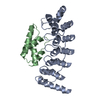


 PDBj
PDBj


 Assembly
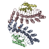


 Mass: 238.305 Da / Num. of mol.: 1 / Source method: obtained synthetically / Formula: C8H18N2O4S / Comment: pH buffer*YM
 Mass: 18.015 Da / Num. of mol.: 233 / Source method: isolated from a natural source / Formula: H2O
 Sample preparation
 / Beamline: BL-6A / Wavelength: 1
  Processing