 Movie
Movie Controller

[English] 日本語
 Yorodumi
- PDB-2yfv: The heterotrimeric complex of Kluyveromyces lactis Scm3, Cse4 and H4 -
+ Open data

- Basic information
| Entry | Database: PDB / ID: 2yfv | ||||||
|---|---|---|---|---|---|---|---|
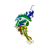
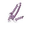
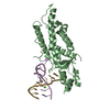
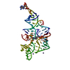
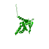
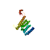
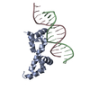
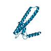
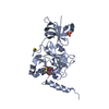
| Title | The heterotrimeric complex of Kluyveromyces lactis Scm3, Cse4 and H4 | ||||||
 Components Components |
| ||||||
 Keywords Keywords | CELL CYCLE / KINETOCHORE / CENTROMERE / HISTONE CHAPERONE / BUDDING YEAST | ||||||
| Function / homology |  Function and homology information Function and homology informationCENP-A containing nucleosome / structural constituent of chromatin / histone binding / protein heterodimerization activity / DNA binding / nucleus Similarity search - Function | ||||||
| Biological species |  KLUYVEROMYCES LACTIS NRRL Y-1140 (yeast) KLUYVEROMYCES LACTIS NRRL Y-1140 (yeast) | ||||||
| Method |  X-RAY DIFFRACTION / SYNCHROTRON / MOLECULAR REPLACEMENT / Resolution: 2.32 Å X-RAY DIFFRACTION / SYNCHROTRON / MOLECULAR REPLACEMENT / Resolution: 2.32 Å | ||||||
 Authors Authors | Cho, U.S. / Harrison, S.C. | ||||||
 Citation Citation | Journal: Proc.Natl.Acad.Sci.USA / Year: 2011 Title: Recognition of the Centromere-Specific Histone Cse4 by the Chaperone Scm3. Authors: Cho, U.S. / Harrison, S.C. | ||||||
| History |
|
- Structure visualization
| Structure viewer | Molecule: MolmilJmol/JSmol |
|---|
- Downloads & links
-Download
| PDBx/mmCIF format | 2yfv.cif.gz | 98.3 KB | Display | PDBx/mmCIF format |
|---|---|---|---|---|
| PDB format | pdb2yfv.ent.gz | 75 KB | Display | PDB format |
| PDBx/mmJSON format | 2yfv.json.gz | Tree view | PDBx/mmJSON format | |
| Others |  Other downloads Other downloads |
-Validation report
| Arichive directory | https://data.pdbj.org/pub/pdb/validation_reports/yf/2yfvftp://data.pdbj.org/pub/pdb/validation_reports/yf/2yfv | HTTPS FTP |
|---|
-Related structure data
| Related structure data | 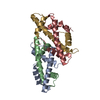 2yfwC 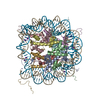 1eqzS C: citing same article ( S: Starting model for refinement |
|---|---|
| Similar structure data |

-Links
 PDBj
PDBj

- Assembly
| Deposited unit | 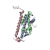
| ||||||||
|---|---|---|---|---|---|---|---|---|---|
| 1 |
| ||||||||
| Unit cell |
|
-Components
| #1: Protein | Mass: 11692.733 Da / Num. of mol.: 1 / Fragment: RESIDUES 81-180 Source method: isolated from a genetically manipulated source Source: (gene. exp.) KLUYVEROMYCES LACTIS NRRL Y-1140 (yeast)Plasmid: PET3A / Production host:  | ||
|---|---|---|---|
| #2: Protein | Mass: 8456.876 Da / Num. of mol.: 1 / Fragment: RESIDUES 25-98 Source method: isolated from a genetically manipulated source Source: (gene. exp.) KLUYVEROMYCES LACTIS NRRL Y-1140 (yeast)Plasmid: PET3A / Production host: | ||
| #3: Protein | Mass: 7430.433 Da / Num. of mol.: 1 / Fragment: RESIDUES 41-103 Source method: isolated from a genetically manipulated source Source: (gene. exp.) KLUYVEROMYCES LACTIS NRRL Y-1140 (yeast)Plasmid: PET3A / Production host: | ||
| #4: Chemical |   Mass: 126.904 Da / Num. of mol.: 2 / Source method: obtained synthetically / Formula: I Mass: 126.904 Da / Num. of mol.: 2 / Source method: obtained synthetically / Formula: I#5: Water | ChemComp-HOH / |  Mass: 18.015 Da / Num. of mol.: 58 / Source method: isolated from a natural source / Formula: H2O Mass: 18.015 Da / Num. of mol.: 58 / Source method: isolated from a natural source / Formula: H2O |
-Experimental details
-Experiment
| Experiment | Method: X-RAY DIFFRACTION / Number of used crystals: 1 |
|---|
- Sample preparation
| Crystal | Density Matthews: 2.59 Å3/Da / Density % sol: 52 % / Description: NONE |
|---|---|
| Crystal grow | pH: 4.5 Details: 0.1 M SODIUM CITRATE, PH4.5, 6% PEG 4000, AND 0.1 M SODIUM IODIDE |
-Data collection
| Diffraction | Mean temperature: 93 K |
|---|---|
| Diffraction source | Source: SYNCHROTRON / Site: APS  / Beamline: 24-ID-E / Wavelength: 0.97914 / Beamline: 24-ID-E / Wavelength: 0.97914 |
| Detector | Type: ADSC QUANTUM 315 / Detector: CCD / Date: Oct 10, 2010 / Details: MIRRORS |
| Radiation | Monochromator: CHOZU HLD8-24 MONOCHROMATOR / Protocol: SINGLE WAVELENGTH / Monochromatic (M) / Laue (L): M / Scattering type: x-ray |
| Radiation wavelength | Wavelength: 0.97914 Å / Relative weight: 1 |
| Reflection | Resolution: 2.3→50 Å / Num. obs: 11636 / % possible obs: 94.7 % / Observed criterion σ(I): 1.5 / Redundancy: 4.1 % / Biso Wilson estimate: 47.7 Å2 / Rmerge(I) obs: 0.08 / Net I/σ(I): 17 |
| Reflection shell | Resolution: 2.3→2.38 Å / Redundancy: 2.8 % / Rmerge(I) obs: 0.64 / Mean I/σ(I) obs: 1.5 / % possible all: 55.3 |
- Processing
| Software |
| ||||||||||||||||||||||||||||||||||||||||||||||||||||||||||||||||||||||||||||||||||||||||||||||||||||||||||||||||||||||||||||||||||||||||||||||||||||||||||||||||||||||||||||||||||||||
|---|---|---|---|---|---|---|---|---|---|---|---|---|---|---|---|---|---|---|---|---|---|---|---|---|---|---|---|---|---|---|---|---|---|---|---|---|---|---|---|---|---|---|---|---|---|---|---|---|---|---|---|---|---|---|---|---|---|---|---|---|---|---|---|---|---|---|---|---|---|---|---|---|---|---|---|---|---|---|---|---|---|---|---|---|---|---|---|---|---|---|---|---|---|---|---|---|---|---|---|---|---|---|---|---|---|---|---|---|---|---|---|---|---|---|---|---|---|---|---|---|---|---|---|---|---|---|---|---|---|---|---|---|---|---|---|---|---|---|---|---|---|---|---|---|---|---|---|---|---|---|---|---|---|---|---|---|---|---|---|---|---|---|---|---|---|---|---|---|---|---|---|---|---|---|---|---|---|---|---|---|---|---|---|
| Refinement | Method to determine structure: MOLECULAR REPLACEMENT Starting model: PDB ENTRY 1EQZ Resolution: 2.32→50 Å / Cor.coef. Fo:Fc: 0.939 / Cor.coef. Fo:Fc free: 0.923 / SU B: 14.225 / SU ML: 0.173 / Cross valid method: THROUGHOUT / ESU R: 0.329 / ESU R Free: 0.239 / Stereochemistry target values: MAXIMUM LIKELIHOOD Details: HYDROGENS HAVE BEEN ADDED IN THE RIDING POSITIONS. RESIDUES 126-130 OF CHAIN A ARE DISORDERED.
| ||||||||||||||||||||||||||||||||||||||||||||||||||||||||||||||||||||||||||||||||||||||||||||||||||||||||||||||||||||||||||||||||||||||||||||||||||||||||||||||||||||||||||||||||||||||
| Solvent computation | Ion probe radii: 0.8 Å / Shrinkage radii: 0.8 Å / VDW probe radii: 1.4 Å / Solvent model: MASK | ||||||||||||||||||||||||||||||||||||||||||||||||||||||||||||||||||||||||||||||||||||||||||||||||||||||||||||||||||||||||||||||||||||||||||||||||||||||||||||||||||||||||||||||||||||||
| Displacement parameters | Biso mean: 50.054 Å2
| ||||||||||||||||||||||||||||||||||||||||||||||||||||||||||||||||||||||||||||||||||||||||||||||||||||||||||||||||||||||||||||||||||||||||||||||||||||||||||||||||||||||||||||||||||||||
| Refinement step | Cycle: LAST / Resolution: 2.32→50 Å
| ||||||||||||||||||||||||||||||||||||||||||||||||||||||||||||||||||||||||||||||||||||||||||||||||||||||||||||||||||||||||||||||||||||||||||||||||||||||||||||||||||||||||||||||||||||||
| Refine LS restraints |
|