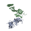
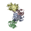
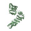
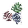
Entry Database : PDB / ID : 5dizTitle Crystal Structure of nuclear proteinaceous RNase P 2 (PRORP2) from A. thaliana Proteinaceous RNase P 2 Keywords / / / / / / / / / Function / homology Function Domain/homology Component
/ / / / / / / / / / / / / / / / / / / / / / / / / / / / Biological species Arabidopsis thaliana (thale cress)Method / / / Resolution : 3.2 Å Authors Karasik, A. / Shanmuganathan, A. / Howard, M.J. / Fierke, C.A. / Koutmos, M. Journal : J.Mol.Biol. / Year : 2016Title : Nuclear Protein-Only Ribonuclease P2 Structure and Biochemical Characterization Provide Insight into the Conserved Properties of tRNA 5' End Processing Enzymes.Authors : Karasik, A. / Shanmuganathan, A. / Howard, M.J. / Fierke, C.A. / Koutmos, M. History Deposition Sep 1, 2015 Deposition site / Processing site Revision 1.0 Dec 30, 2015 Provider / Type Revision 1.1 Feb 17, 2016 Group Revision 1.2 Nov 1, 2017 Group / Database references / Derived calculationsCategory / pdbx_struct_assembly_auth_evidence / pdbx_struct_oper_listItem / _pdbx_struct_oper_list.symmetry_operationRevision 1.3 Sep 27, 2023 Group / Database references / Refinement descriptionCategory chem_comp_atom / chem_comp_bond ... chem_comp_atom / chem_comp_bond / database_2 / pdbx_initial_refinement_model Item / _database_2.pdbx_database_accession
Show all Show less
 Movie
Movie Controller

 Yorodumi
 Open data
 Basic information
 Components
 Keywords
 Function and homology information
 X-RAY DIFFRACTION /
  Authors
 Citation
 Structure visualization
 Downloads & links
 Other downloads
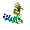


 PDBj
PDBj
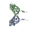
 Assembly
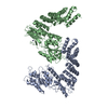


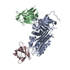
 Mass: 65.409 Da / Num. of mol.: 2 / Source method: obtained synthetically / Formula: Zn
 Mass: 18.015 Da / Num. of mol.: 5 / Source method: isolated from a natural source / Formula: H2O
 Sample preparation
 / Beamline: 23-ID-B / Wavelength: 1.033 Å
 Processing