 Movie
Movie Controller

[English] 日本語
 Yorodumi
- PDB-6gwp: Crystal Structure of Stabilized Active Plasminogen Activator Inhi... -
+ Open data

- Basic information
| Entry | Database: PDB / ID: 6gwp | |||||||||
|---|---|---|---|---|---|---|---|---|---|---|
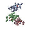
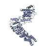
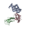
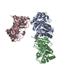
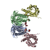
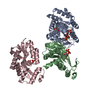
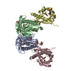
| Title | Crystal Structure of Stabilized Active Plasminogen Activator Inhibitor-1 (PAI-1-stab) in Complex with Two Inhibitory Nanobodies (VHH-2g-42, VHH-2w-64) | |||||||||
 Components Components |
| |||||||||
 Keywords Keywords | HYDROLASE / plasminogen activator inhibitor-1 / PAI-1 / PAI-1-stab / serpin / protease inhibitor / serine protease inhibitor / nanobody / antibody fragment / protein complex | |||||||||
| Function / homology |  Function and homology information Function and homology informationpositive regulation of leukotriene production involved in inflammatory response / negative regulation of smooth muscle cell-matrix adhesion / negative regulation of integrin-mediated signaling pathway / peptidase inhibitor complex / dentinogenesis / positive regulation of coagulation / negative regulation of vascular wound healing / negative regulation of smooth muscle cell migration / Regulation of MITF-M-dependent genes involved in extracellular matrix, focal adhesion and epithelial-to-mesenchymal transition / negative regulation of wound healing ...positive regulation of leukotriene production involved in inflammatory response / negative regulation of smooth muscle cell-matrix adhesion / negative regulation of integrin-mediated signaling pathway / peptidase inhibitor complex / dentinogenesis / positive regulation of coagulation / negative regulation of vascular wound healing / negative regulation of smooth muscle cell migration / Regulation of MITF-M-dependent genes involved in extracellular matrix, focal adhesion and epithelial-to-mesenchymal transition / negative regulation of wound healing / positive regulation of odontoblast differentiation / negative regulation of plasminogen activation / Dissolution of Fibrin Clot / negative regulation of cell adhesion mediated by integrin / positive regulation of monocyte chemotaxis / endopeptidase inhibitor activity / negative regulation of thrombin-activated receptor signaling pathway / negative regulation of blood coagulation / negative regulation of fibrinolysis / positive regulation of blood coagulation / replicative senescence / ECM proteoglycans / negative regulation of endothelial cell apoptotic process / negative regulation of extrinsic apoptotic signaling pathway via death domain receptors / serine protease inhibitor complex / fibrinolysis / negative regulation of proteolysis / BMAL1:CLOCK,NPAS2 activates circadian expression / platelet alpha granule lumen / negative regulation of cell migration / positive regulation of interleukin-8 production / serine-type endopeptidase inhibitor activity / SMAD2/SMAD3:SMAD4 heterotrimer regulates transcription / positive regulation of receptor-mediated endocytosis / positive regulation of angiogenesis / positive regulation of inflammatory response / Platelet degranulation / extracellular matrix / cellular response to lipopolysaccharide / protease binding / angiogenesis / defense response to Gram-negative bacterium / signaling receptor binding / : / extracellular exosome / extracellular region / plasma membrane Similarity search - Function | |||||||||
| Biological species |  Homo sapiens (human) Homo sapiens (human) | |||||||||
| Method |  X-RAY DIFFRACTION / SYNCHROTRON / MOLECULAR REPLACEMENT / molecular replacement / Resolution: 2.28 Å X-RAY DIFFRACTION / SYNCHROTRON / MOLECULAR REPLACEMENT / molecular replacement / Resolution: 2.28 Å | |||||||||
 Authors Authors | Sillen, M. / Weeks, S.D. / Strelkov, S.V. / Declerck, P.J. | |||||||||
| Funding support |  Belgium, 2items Belgium, 2items
| |||||||||
 Citation Citation | Journal: J.Thromb.Haemost. / Year: 2020 Title: Molecular mechanism of two nanobodies that inhibit PAI-1 activity reveals a modulation at distinct stages of the PAI-1/plasminogen activator interaction. Authors: Sillen, M. / Weeks, S.D. / Zhou, X. / Komissarov, A.A. / Florova, G. / Idell, S. / Strelkov, S.V. / Declerck, P.J. | |||||||||
| History |
|
- Structure visualization
| Structure viewer | Molecule: MolmilJmol/JSmol |
|---|
- Downloads & links
-Download
| PDBx/mmCIF format | 6gwp.cif.gz | 250 KB | Display | PDBx/mmCIF format |
|---|---|---|---|---|
| PDB format | pdb6gwp.ent.gz | 201 KB | Display | PDB format |
| PDBx/mmJSON format | 6gwp.json.gz | Tree view | PDBx/mmJSON format | |
| Others |  Other downloads Other downloads |
-Validation report
| Arichive directory | https://data.pdbj.org/pub/pdb/validation_reports/gw/6gwpftp://data.pdbj.org/pub/pdb/validation_reports/gw/6gwp | HTTPS FTP |
|---|
-Related structure data
| Related structure data | 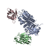 6gwnC 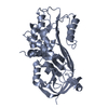 6gwqC  1db2S 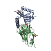 5ja8S 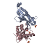 5ja9S S: Starting model for refinement C: citing same article ( |
|---|---|
| Similar structure data |


-Links
 PDBj
PDBj


- Assembly
| Deposited unit | 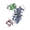
| ||||||||
|---|---|---|---|---|---|---|---|---|---|
| 1 |
| ||||||||
| Unit cell |
|
-Components
| #1: Protein | Mass: 42751.008 Da / Num. of mol.: 1 / Mutation: N150H-K154T-Q301P-Q319L-M354I Source method: isolated from a genetically manipulated source Source: (gene. exp.) Homo sapiens (human) / Plasmid: pETHSUK2 / Production host:  |
|---|---|
| #2: Antibody | Mass: 12752.254 Da / Num. of mol.: 1 Source method: isolated from a genetically manipulated source Source: (gene. exp.) |
| #3: Antibody | Mass: 13087.387 Da / Num. of mol.: 1 Source method: isolated from a genetically manipulated source Source: (gene. exp.) |
| #4: Water | ChemComp-HOH /  Mass: 18.015 Da / Num. of mol.: 64 / Source method: isolated from a natural source / Formula: H2O Mass: 18.015 Da / Num. of mol.: 64 / Source method: isolated from a natural source / Formula: H2O |
| Has protein modification | Y |
-Experimental details
-Experiment
| Experiment | Method: X-RAY DIFFRACTION / Number of used crystals: 1 |
|---|
- Sample preparation
| Crystal | Density Matthews: 2.2 Å3/Da / Density % sol: 44.14 % / Mosaicity: 0.17 ° |
|---|---|
| Crystal grow | Temperature: 293 K / Method: vapor diffusion, sitting drop / pH: 6.5 Details: 0.1 M Bis-Tris, 17 % w/v PEG 3350, 3 % v/v methanol |
-Data collection
| Diffraction | Mean temperature: 100 K | ||||||||||||||||||||||||||||||
|---|---|---|---|---|---|---|---|---|---|---|---|---|---|---|---|---|---|---|---|---|---|---|---|---|---|---|---|---|---|---|---|
| Diffraction source | Source: SYNCHROTRON / Site: ESRF  / Beamline: MASSIF-3 / Wavelength: 0.9677 Å / Beamline: MASSIF-3 / Wavelength: 0.9677 Å | ||||||||||||||||||||||||||||||
| Detector | Type: DECTRIS EIGER X 4M / Detector: PIXEL / Date: Jul 21, 2017 Details: CRLs and a half-Kirkpatrick-Baez (KB) geometry as the vertical and horizontal focusing systems | ||||||||||||||||||||||||||||||
| Radiation | Monochromator: Si (111) monochromator / Protocol: SINGLE WAVELENGTH / Monochromatic (M) / Laue (L): M / Scattering type: x-ray | ||||||||||||||||||||||||||||||
| Radiation wavelength | Wavelength: 0.9677 Å / Relative weight: 1 | ||||||||||||||||||||||||||||||
| Reflection | Resolution: 2.18→97.674 Å / Num. obs: 30312 / % possible obs: 97.5 % / Redundancy: 3.8 % / Biso Wilson estimate: 46.45 Å2 / CC1/2: 0.993 / Rmerge(I) obs: 0.102 / Rpim(I) all: 0.062 / Rrim(I) all: 0.119 / Net I/σ(I): 9.2 / Num. measured all: 115963 | ||||||||||||||||||||||||||||||
| Reflection shell | Diffraction-ID: 1
|
-Phasing
| Phasing | Method: molecular replacement |
|---|
- Processing
| Software |
| ||||||||||||||||||||||||||||||||||||||||||||||||||||||||||||||||||||||||||||||||||||||||||||||||||||
|---|---|---|---|---|---|---|---|---|---|---|---|---|---|---|---|---|---|---|---|---|---|---|---|---|---|---|---|---|---|---|---|---|---|---|---|---|---|---|---|---|---|---|---|---|---|---|---|---|---|---|---|---|---|---|---|---|---|---|---|---|---|---|---|---|---|---|---|---|---|---|---|---|---|---|---|---|---|---|---|---|---|---|---|---|---|---|---|---|---|---|---|---|---|---|---|---|---|---|---|---|---|
| Refinement | Method to determine structure: MOLECULAR REPLACEMENT Starting model: 1DB2, 5JA8, 5JA9 Resolution: 2.28→97.674 Å / SU ML: 0.33 / Cross valid method: THROUGHOUT / σ(F): 0.31 / Phase error: 28.89 / Stereochemistry target values: ML
| ||||||||||||||||||||||||||||||||||||||||||||||||||||||||||||||||||||||||||||||||||||||||||||||||||||
| Solvent computation | Shrinkage radii: 0.9 Å / VDW probe radii: 1.11 Å / Solvent model: FLAT BULK SOLVENT MODEL | ||||||||||||||||||||||||||||||||||||||||||||||||||||||||||||||||||||||||||||||||||||||||||||||||||||
| Displacement parameters | Biso max: 138.83 Å2 / Biso mean: 61.2692 Å2 / Biso min: 27.51 Å2 | ||||||||||||||||||||||||||||||||||||||||||||||||||||||||||||||||||||||||||||||||||||||||||||||||||||
| Refinement step | Cycle: final / Resolution: 2.28→97.674 Å
| ||||||||||||||||||||||||||||||||||||||||||||||||||||||||||||||||||||||||||||||||||||||||||||||||||||
| Refine LS restraints |
| ||||||||||||||||||||||||||||||||||||||||||||||||||||||||||||||||||||||||||||||||||||||||||||||||||||
| LS refinement shell | Refine-ID: X-RAY DIFFRACTION / Rfactor Rfree error: 0 / Total num. of bins used: 10
| ||||||||||||||||||||||||||||||||||||||||||||||||||||||||||||||||||||||||||||||||||||||||||||||||||||
| Refinement TLS params. | Method: refined / Refine-ID: X-RAY DIFFRACTION
| ||||||||||||||||||||||||||||||||||||||||||||||||||||||||||||||||||||||||||||||||||||||||||||||||||||
| Refinement TLS group |
|
