 Movie
Movie Controller


+ Open data

- Basic information
| Entry | Database: PDB / ID: 5dgo | ||||||
|---|---|---|---|---|---|---|---|
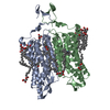
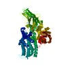
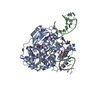
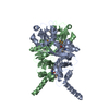
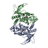
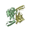
| Title | Crystal structure of cell division cycle protein 45 (Cdc45) | ||||||
 Components Components | Cell division control protein 45 homolog | ||||||
 Keywords Keywords | CELL CYCLE / DNA replication / CMG helicase subunit / RecJ fold | ||||||
| Function / homology |  Function and homology information Function and homology informationUnwinding of DNA / mitotic DNA replication preinitiation complex assembly / CMG complex / DNA replication checkpoint signaling / DNA replication preinitiation complex / double-strand break repair via break-induced replication / G1/S-Specific Transcription / DNA unwinding involved in DNA replication / DNA replication origin binding / Activation of the pre-replicative complex ...Unwinding of DNA / mitotic DNA replication preinitiation complex assembly / CMG complex / DNA replication checkpoint signaling / DNA replication preinitiation complex / double-strand break repair via break-induced replication / G1/S-Specific Transcription / DNA unwinding involved in DNA replication / DNA replication origin binding / Activation of the pre-replicative complex / DNA replication initiation / Activation of ATR in response to replication stress / ciliary basal body / single-stranded DNA binding / centrosome / chromatin binding / nucleoplasm / nucleus Similarity search - Function | ||||||
| Biological species |  Homo sapiens (human) Homo sapiens (human) | ||||||
| Method |  X-RAY DIFFRACTION / SYNCHROTRON / SAD / Resolution: 2.1 Å X-RAY DIFFRACTION / SYNCHROTRON / SAD / Resolution: 2.1 Å | ||||||
 Authors Authors | Simon, A.C. / Pellegrini, L. | ||||||
| Funding support |  United Kingdom, 1items United Kingdom, 1items
| ||||||
 Citation Citation | Journal: Nat Commun / Year: 2016 Title: Structure of human Cdc45 and implications for CMG helicase function. Authors: Simon, A.C. / Sannino, V. / Costanzo, V. / Pellegrini, L. | ||||||
| History |
|
- Structure visualization
| Structure viewer | Molecule: MolmilJmol/JSmol |
|---|
- Downloads & links
-Download
| PDBx/mmCIF format | 5dgo.cif.gz | 235.1 KB | Display | PDBx/mmCIF format |
|---|---|---|---|---|
| PDB format | pdb5dgo.ent.gz | 198.9 KB | Display | PDB format |
| PDBx/mmJSON format | 5dgo.json.gz | Tree view | PDBx/mmJSON format | |
| Others |  Other downloads Other downloads |
-Validation report
| Summary document | 5dgo_validation.pdf.gz | 441.2 KB | Display | wwPDB validaton report |
|---|---|---|---|---|
| Full document | 5dgo_full_validation.pdf.gz | 441.6 KB | Display | |
| Data in XML | 5dgo_validation.xml.gz | 26.2 KB | Display | |
| Data in CIF | 5dgo_validation.cif.gz | 40.1 KB | Display | |
| Arichive directory | https://data.pdbj.org/pub/pdb/validation_reports/dg/5dgoftp://data.pdbj.org/pub/pdb/validation_reports/dg/5dgo | HTTPS FTP |
-Related structure data
| Similar structure data |
|---|


-Links
 PDBj
PDBj


- Assembly
| Deposited unit | 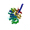
| ||||||||
|---|---|---|---|---|---|---|---|---|---|
| 1 |
| ||||||||
| Unit cell |
|
-Components
| #1: Protein | Mass: 64277.008 Da / Num. of mol.: 1 Source method: isolated from a genetically manipulated source Details: Residues 154 to 164 (11 aminoacids) were left out of the expression construct used for determination of the crystal structure. Source: (gene. exp.) Homo sapiens (human) / Gene: CDC45, CDC45L, CDC45L2, UNQ374/PRO710 / Plasmid: pRSFDuet-1 / Production host:  | ||||
|---|---|---|---|---|---|
| #2: Chemical | ChemComp-EDO /   Mass: 62.068 Da / Num. of mol.: 6 / Source method: obtained synthetically / Formula: C2H6O2 Mass: 62.068 Da / Num. of mol.: 6 / Source method: obtained synthetically / Formula: C2H6O2#3: Chemical |   Mass: 96.063 Da / Num. of mol.: 3 / Source method: obtained synthetically / Formula: SO4 Mass: 96.063 Da / Num. of mol.: 3 / Source method: obtained synthetically / Formula: SO4#4: Water | ChemComp-HOH / |  Mass: 18.015 Da / Num. of mol.: 541 / Source method: isolated from a natural source / Formula: H2O Mass: 18.015 Da / Num. of mol.: 541 / Source method: isolated from a natural source / Formula: H2O |
-Experimental details
-Experiment
| Experiment | Method: X-RAY DIFFRACTION |
|---|
- Sample preparation
| Crystal | Density Matthews: 4.09 Å3/Da / Density % sol: 69.91 % |
|---|---|
| Crystal grow | Temperature: 292 K / Method: vapor diffusion, sitting drop Details: Buffer condition C2 of the Morpheus HT 96 crystallisation screen (Molecular Dimensions) |
-Data collection
| Diffraction | Mean temperature: 100 K |
|---|---|
| Diffraction source | Source: SYNCHROTRON / Site: Diamond / Beamline: I24 / Wavelength: 0.9778 Å |
| Detector | Type: DECTRIS PILATUS 6M / Detector: PIXEL / Date: May 28, 2013 |
| Radiation | Protocol: SINGLE WAVELENGTH / Monochromatic (M) / Laue (L): M / Scattering type: x-ray |
| Radiation wavelength | Wavelength: 0.9778 Å / Relative weight: 1 |
| Reflection | Resolution: 2.1→48.84 Å / Num. obs: 61965 / % possible obs: 99.8 % / Redundancy: 6.6 % / Biso Wilson estimate: 33.7 Å2 / Net I/σ(I): 11.8 |
| Reflection shell | Resolution: 2.1→2.15 Å / Redundancy: 6.5 % / Rmerge(I) obs: 1.042 / Mean I/σ(I) obs: 2.1 / % possible all: 97.1 |
- Processing
| Software |
| |||||||||||||||||||||||||||||||||||||||||||||||||||||||||||||||||||||||||||||||||||||||||||||||||||||||||||||||||||||||||||||||||||||||||||||||||||||||||||||||||||||||||||||||||||||||||||||||||||||||||||||||||||||||||
|---|---|---|---|---|---|---|---|---|---|---|---|---|---|---|---|---|---|---|---|---|---|---|---|---|---|---|---|---|---|---|---|---|---|---|---|---|---|---|---|---|---|---|---|---|---|---|---|---|---|---|---|---|---|---|---|---|---|---|---|---|---|---|---|---|---|---|---|---|---|---|---|---|---|---|---|---|---|---|---|---|---|---|---|---|---|---|---|---|---|---|---|---|---|---|---|---|---|---|---|---|---|---|---|---|---|---|---|---|---|---|---|---|---|---|---|---|---|---|---|---|---|---|---|---|---|---|---|---|---|---|---|---|---|---|---|---|---|---|---|---|---|---|---|---|---|---|---|---|---|---|---|---|---|---|---|---|---|---|---|---|---|---|---|---|---|---|---|---|---|---|---|---|---|---|---|---|---|---|---|---|---|---|---|---|---|---|---|---|---|---|---|---|---|---|---|---|---|---|---|---|---|---|---|---|---|---|---|---|---|---|---|---|---|---|---|---|---|---|
| Refinement | Method to determine structure: SAD / Resolution: 2.1→48.84 Å / SU ML: 0.22 / Cross valid method: FREE R-VALUE / Phase error: 20.07 / Stereochemistry target values: ML
| |||||||||||||||||||||||||||||||||||||||||||||||||||||||||||||||||||||||||||||||||||||||||||||||||||||||||||||||||||||||||||||||||||||||||||||||||||||||||||||||||||||||||||||||||||||||||||||||||||||||||||||||||||||||||
| Solvent computation | Shrinkage radii: 0.9 Å / VDW probe radii: 1.11 Å / Solvent model: FLAT BULK SOLVENT MODEL | |||||||||||||||||||||||||||||||||||||||||||||||||||||||||||||||||||||||||||||||||||||||||||||||||||||||||||||||||||||||||||||||||||||||||||||||||||||||||||||||||||||||||||||||||||||||||||||||||||||||||||||||||||||||||
| Displacement parameters | Biso mean: 43.8 Å2 | |||||||||||||||||||||||||||||||||||||||||||||||||||||||||||||||||||||||||||||||||||||||||||||||||||||||||||||||||||||||||||||||||||||||||||||||||||||||||||||||||||||||||||||||||||||||||||||||||||||||||||||||||||||||||
| Refinement step | Cycle: LAST / Resolution: 2.1→48.84 Å
| |||||||||||||||||||||||||||||||||||||||||||||||||||||||||||||||||||||||||||||||||||||||||||||||||||||||||||||||||||||||||||||||||||||||||||||||||||||||||||||||||||||||||||||||||||||||||||||||||||||||||||||||||||||||||
| Refine LS restraints |
| |||||||||||||||||||||||||||||||||||||||||||||||||||||||||||||||||||||||||||||||||||||||||||||||||||||||||||||||||||||||||||||||||||||||||||||||||||||||||||||||||||||||||||||||||||||||||||||||||||||||||||||||||||||||||
| LS refinement shell |
|
