 Movie
Movie Controller

[English] 日本語
 Yorodumi
- PDB-6ezd: Pyrrolysyl-tRNA synthetase from Canditatus Methanomethylophilus a... -
+ Open data

- Basic information
| Entry | Database: PDB / ID: 6ezd | ||||||
|---|---|---|---|---|---|---|---|
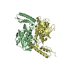
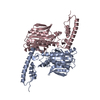
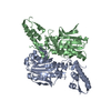
| Title | Pyrrolysyl-tRNA synthetase from Canditatus Methanomethylophilus alvus (MmaPylRS) | ||||||
 Components Components | Pyrrolysyl-tRNA synthetase | ||||||
 Keywords Keywords | LIGASE / Pyrrolysyl-tRNA synthetase | ||||||
| Function / homology |  Function and homology information Function and homology informationpyrrolysine-tRNAPyl ligase / pyrrolysyl-tRNA synthetase activity / metal ion binding Similarity search - Function | ||||||
| Biological species |  Candidatus Methanomethylophilus alvus Mx1201 1) (archaea) Candidatus Methanomethylophilus alvus Mx1201 1) (archaea) | ||||||
| Method |  X-RAY DIFFRACTION / SYNCHROTRON / MOLECULAR REPLACEMENT / Resolution: 2.4 Å X-RAY DIFFRACTION / SYNCHROTRON / MOLECULAR REPLACEMENT / Resolution: 2.4 Å | ||||||
 Authors Authors | Pavkov-Keller, T. / Schweiger, K. / Gruber, K. | ||||||
| Funding support |  Austria, 1items Austria, 1items
| ||||||
 Citation Citation | Journal: to be published Title: A new archaeal pyrrolysyl-tRNA synthetase/amber suppressor tRNA pair for orthogonal protein translation Authors: Fladischer, P. / Blamauer, J. / Pavkov-Keller, T. / Schweiger, K. / Darnhofer, B. / Birner-Gruenberger, R. / Gruber, K. / Wiltschi, B. | ||||||
| History |
|
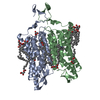
- Structure visualization
| Structure viewer | Molecule: MolmilJmol/JSmol |
|---|
- Downloads & links
-Download
| PDBx/mmCIF format | 6ezd.cif.gz | 226.5 KB | Display | PDBx/mmCIF format |
|---|---|---|---|---|
| PDB format | pdb6ezd.ent.gz | 183.8 KB | Display | PDB format |
| PDBx/mmJSON format | 6ezd.json.gz | Tree view | PDBx/mmJSON format | |
| Others |  Other downloads Other downloads |
-Validation report
| Arichive directory | https://data.pdbj.org/pub/pdb/validation_reports/ez/6ezdftp://data.pdbj.org/pub/pdb/validation_reports/ez/6ezd | HTTPS FTP |
|---|
-Related structure data
| Related structure data | 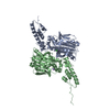 3dsqS S: Starting model for refinement |
|---|---|
| Similar structure data |

-Links
 PDBj
PDBj
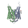
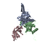
- Assembly
| Deposited unit | 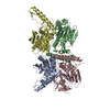
| ||||||||
|---|---|---|---|---|---|---|---|---|---|
| 1 | 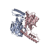
| ||||||||
| 2 | 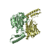
| ||||||||
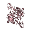
| Unit cell |
| ||||||||
| Components on special symmetry positions |
|
-Components
| #1: Protein | Mass: 31819.096 Da / Num. of mol.: 4 Source method: isolated from a genetically manipulated source Source: (gene. exp.) Candidatus Methanomethylophilus alvus Mx1201 1) (archaea)Gene: MMALV_11280 / Production host:  #2: Water | ChemComp-HOH / |  Mass: 18.015 Da / Num. of mol.: 364 / Source method: isolated from a natural source / Formula: H2O Mass: 18.015 Da / Num. of mol.: 364 / Source method: isolated from a natural source / Formula: H2O |
|---|
-Experimental details
-Experiment
| Experiment | Method: X-RAY DIFFRACTION / Number of used crystals: 1 |
|---|
- Sample preparation
| Crystal | Density Matthews: 2.48 Å3/Da / Density % sol: 50.41 % |
|---|---|
| Crystal grow | Temperature: 289 K / Method: vapor diffusion, sitting drop Details: 0.09 M (NaNO3, Na2HPO4, (NH4)2SO4), 0.1 M Tris (base), bicine and 5 mM MgCl2. 15-7mg/ml (in 10 mM HEPES-Na pH 7.4, 300 mM NaCl) |
-Data collection
| Diffraction | Mean temperature: 100 K |
|---|---|
| Diffraction source | Source: SYNCHROTRON / Site: PETRA III, DESY  / Beamline: P11 / Wavelength: 0.9794 Å / Beamline: P11 / Wavelength: 0.9794 Å |
| Detector | Type: DECTRIS PILATUS 6M / Detector: PIXEL / Date: Jul 18, 2016 |
| Radiation | Protocol: SINGLE WAVELENGTH / Monochromatic (M) / Laue (L): M / Scattering type: x-ray |
| Radiation wavelength | Wavelength: 0.9794 Å / Relative weight: 1 |
| Reflection | Resolution: 2.4→48.78 Å / Num. obs: 49665 / % possible obs: 99 % / Redundancy: 6.4 % / Biso Wilson estimate: 37.17 Å2 / CC1/2: 0.996 / Rmerge(I) obs: 0.117 / Rpim(I) all: 0.05 / Rrim(I) all: 0.128 / Net I/σ(I): 13.04 |
| Reflection shell | Resolution: 2.4→2.49 Å / Redundancy: 6.6 % / Rmerge(I) obs: 0.664 / Num. unique obs: 4856 / CC1/2: 0.772 / Rpim(I) all: 0.277 / Rrim(I) all: 0.721 / % possible all: 98 |
- Processing
| Software |
| |||||||||||||||||||||||||||||||||||||||||||||||||||||||||||||||||||||||||||||||||||||||||||||||||||||||||||||||||||||||||||||||||||||
|---|---|---|---|---|---|---|---|---|---|---|---|---|---|---|---|---|---|---|---|---|---|---|---|---|---|---|---|---|---|---|---|---|---|---|---|---|---|---|---|---|---|---|---|---|---|---|---|---|---|---|---|---|---|---|---|---|---|---|---|---|---|---|---|---|---|---|---|---|---|---|---|---|---|---|---|---|---|---|---|---|---|---|---|---|---|---|---|---|---|---|---|---|---|---|---|---|---|---|---|---|---|---|---|---|---|---|---|---|---|---|---|---|---|---|---|---|---|---|---|---|---|---|---|---|---|---|---|---|---|---|---|---|---|---|
| Refinement | Method to determine structure: MOLECULAR REPLACEMENT Starting model: 3DSQ Resolution: 2.4→48.78 Å / SU ML: 0.32 / Cross valid method: FREE R-VALUE / σ(F): 1.35 / Phase error: 26.42
| |||||||||||||||||||||||||||||||||||||||||||||||||||||||||||||||||||||||||||||||||||||||||||||||||||||||||||||||||||||||||||||||||||||
| Solvent computation | Shrinkage radii: 0.9 Å / VDW probe radii: 1.11 Å | |||||||||||||||||||||||||||||||||||||||||||||||||||||||||||||||||||||||||||||||||||||||||||||||||||||||||||||||||||||||||||||||||||||
| Refinement step | Cycle: LAST / Resolution: 2.4→48.78 Å
| |||||||||||||||||||||||||||||||||||||||||||||||||||||||||||||||||||||||||||||||||||||||||||||||||||||||||||||||||||||||||||||||||||||
| Refine LS restraints |
| |||||||||||||||||||||||||||||||||||||||||||||||||||||||||||||||||||||||||||||||||||||||||||||||||||||||||||||||||||||||||||||||||||||
| LS refinement shell |
|
