 Movie
Movie Controller


+ Open data

- Basic information
| Entry | Database: PDB / ID: 5osg | |||||||||
|---|---|---|---|---|---|---|---|---|---|---|
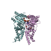
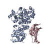
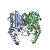
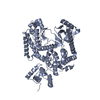
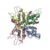
| Title | Structure of KSRP in context of Leishmania donovani 80S | |||||||||
 Components Components |
| |||||||||
 Keywords Keywords | RIBOSOME / Kinetoplastids / cryo-EM / KSRP | |||||||||
| Function / homology |  Function and homology information Function and homology informationstructural constituent of ribosome / ribosome / translation / ribonucleoprotein complex / RNA binding Similarity search - Function | |||||||||
| Biological species |  Leishmania donovani (eukaryote) Leishmania donovani (eukaryote) | |||||||||
| Method | ELECTRON MICROSCOPY / single particle reconstruction / cryo EM / Resolution: 2.9 Å | |||||||||
 Authors Authors | Brito Querido, J. / Mancera-Martinez, E. / Vicens, Q. / Bochler, A. / Chicher, J. / Simonetti, A. / Hashem, Y. | |||||||||
| Funding support |  France, 2items France, 2items
| |||||||||
 Citation Citation | Journal: Structure / Year: 2017 Title: The cryo-EM Structure of a Novel 40S Kinetoplastid-Specific Ribosomal Protein. Authors: Jailson Brito Querido / Eder Mancera-Martínez / Quentin Vicens / Anthony Bochler / Johana Chicher / Angelita Simonetti / Yaser Hashem / Abstract: Kinetoplastids are potentially lethal protozoan pathogens affecting more than 20 million people worldwide. There is a critical need for more specific targets for the development of safer anti- ...Kinetoplastids are potentially lethal protozoan pathogens affecting more than 20 million people worldwide. There is a critical need for more specific targets for the development of safer anti-kinetoplastid therapeutic molecules that can replace the scarce and highly cytotoxic current drugs. The kinetoplastid ribosome represents a potential therapeutic target due to its relative structural divergence when compared with its human counterpart. However, several kinetoplastid-specific ribosomal features remain uncharacterized. Here, we present the near-atomic cryoelectron microscopy structure of a novel bona fide kinetoplastid-specific ribosomal (r-) protein (KSRP) bound to the ribosome. KSRP is an essential protein located at the solvent face of the 40S subunit, where it binds and stabilizes kinetoplastid-specific domains of rRNA, suggesting its role in ribosome integrity. KSRP also interacts with the r-protein eS6 at a region that is only conserved in kinetoplastids. The kinetoplastid-specific ribosomal environment of KSRP provides a promising target for the design of safer anti-kinetoplastidian drugs. #1: Journal: Nat Commun / Year: 2016Title: Structures and stabilization of kinetoplastid-specific split rRNAs revealed by comparing leishmanial and human ribosomes. Authors: Xing Zhang / Mason Lai / Winston Chang / Iris Yu / Ke Ding / Jan Mrazek / Hwee L Ng / Otto O Yang / Dmitri A Maslov / Z Hong Zhou /   Abstract: The recent success in ribosome structure determination by cryoEM has opened the door to defining structural differences between ribosomes of pathogenic organisms and humans and to understand ribosome- ...The recent success in ribosome structure determination by cryoEM has opened the door to defining structural differences between ribosomes of pathogenic organisms and humans and to understand ribosome-targeting antibiotics. Here, by direct electron-counting cryoEM, we have determined the structures of the Leishmania donovani and human ribosomes at 2.9 Å and 3.6 Å, respectively. Our structure of the leishmanial ribosome elucidates the organization of the six fragments of its large subunit rRNA (as opposed to a single 28S rRNA in most eukaryotes, including humans) and reveals atomic details of a unique 20 amino acid extension of the uL13 protein that pins down the ends of three of the rRNA fragments. The structure also fashions many large rRNA expansion segments. Direct comparison of our human and leishmanial ribosome structures at the decoding A-site sheds light on how the bacterial ribosome-targeting drug paromomycin selectively inhibits the eukaryotic L. donovani, but not human, ribosome. | |||||||||
| History |
|
- Structure visualization
| Movie |
Movie viewer |
|---|---|
| Structure viewer | Molecule: MolmilJmol/JSmol |
- Downloads & links
-Download
| PDBx/mmCIF format | 5osg.cif.gz | 138.6 KB | Display | PDBx/mmCIF format |
|---|---|---|---|---|
| PDB format | pdb5osg.ent.gz | 74.3 KB | Display | PDB format |
| PDBx/mmJSON format | 5osg.json.gz | Tree view | PDBx/mmJSON format | |
| Others |  Other downloads Other downloads |
-Validation report
| Arichive directory | https://data.pdbj.org/pub/pdb/validation_reports/os/5osgftp://data.pdbj.org/pub/pdb/validation_reports/os/5osg | HTTPS FTP |
|---|
-Related structure data
| Related structure data | 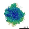 8343M 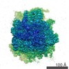 3844C 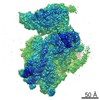 3845C  5optC M: map data used to model this data C: citing same article ( |
|---|---|
| Similar structure data |


-Links
 PDBj
PDBj


- Assembly
| Deposited unit | 
|
|---|---|
| 1 |
|
-Components
| #1: Protein | Mass: 25254.348 Da / Num. of mol.: 1 Source method: isolated from a genetically manipulated source Source: (gene. exp.) Leishmania donovani (eukaryote) / Gene: LDBPK_320790 / Production host: Leishmania donovani (eukaryote) / References: UniProt: E9BNI3 |
|---|---|
| #2: RNA chain | Mass: 710272.000 Da / Num. of mol.: 1 Source method: isolated from a genetically manipulated source Source: (gene. exp.) Leishmania donovani (eukaryote) / Production host: Leishmania donovani (eukaryote) / References: GenBank: 9504 |
| #3: Protein | Mass: 28370.227 Da / Num. of mol.: 1 Source method: isolated from a genetically manipulated source Source: (gene. exp.) Leishmania donovani (eukaryote) / Gene: RPS6, L3640.11, Lmj_1130, LmjF21.1780, LmjF_21_1780 / Production host: Leishmania donovani (eukaryote) / References: UniProt: Q9NE83 |
-Experimental details
-Experiment
| Experiment | Method: ELECTRON MICROSCOPY |
|---|---|
| EM experiment | Aggregation state: PARTICLE / 3D reconstruction method: single particle reconstruction |
- Sample preparation
| Component | Name: 80S ribosome / Type: RIBOSOME / Entity ID: all / Source: NATURAL |
|---|---|
| Source (natural) | Organism: Leishmania donovani (eukaryote) |
| Buffer solution | pH: 7.6 |
| Specimen | Embedding applied: NO / Shadowing applied: NO / Staining applied: NO / Vitrification applied: YES |
| Vitrification | Cryogen name: ETHANE / Humidity: 100 % |
- Electron microscopy imaging
| Experimental equipment |  Model: Titan Krios / Image courtesy: FEI Company |
|---|---|
| Microscopy | Model: FEI TITAN KRIOS |
| Electron gun | Electron source:  FIELD EMISSION GUN / Accelerating voltage: 300 kV / Illumination mode: FLOOD BEAM FIELD EMISSION GUN / Accelerating voltage: 300 kV / Illumination mode: FLOOD BEAM |
| Electron lens | Mode: BRIGHT FIELD |
| Image recording | Electron dose: 20 e/Å2 / Film or detector model: GATAN K2 BASE (4k x 4k) |
- Processing
| CTF correction | Type: PHASE FLIPPING AND AMPLITUDE CORRECTION |
|---|---|
| 3D reconstruction | Resolution: 2.9 Å / Resolution method: FSC 0.143 CUT-OFF / Num. of particles: 213108 / Symmetry type: POINT |
