 Movie
Movie Controller

[English] 日本語
 Yorodumi
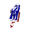
- PDB-5zih: Crystal structure of the red light-activated channelrhodopsin Chr... -
+ Open data

- Basic information
| Entry | Database: PDB / ID: 5zih | |||||||||
|---|---|---|---|---|---|---|---|---|---|---|
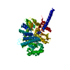
| Title | Crystal structure of the red light-activated channelrhodopsin Chrimson. | |||||||||
 Components Components | Sensory opsin A,Chrimson | |||||||||
 Keywords Keywords | MEMBRANE PROTEIN / rhodopsin / ion channel | |||||||||
| Function / homology | Bacteriorhodopsin-like protein / Archaeal/bacterial/fungal rhodopsins / Bacteriorhodopsin-like protein / photoreceptor activity / phototransduction / membrane / metal ion binding / (2R)-2,3-dihydroxypropyl (9Z)-octadec-9-enoate / Sensory opsin A Function and homology information Function and homology information | |||||||||
| Biological species |   Chlamydomonas reinhardtii (plant)Chlamydomonas noctigama (plant) Chlamydomonas reinhardtii (plant)Chlamydomonas noctigama (plant) | |||||||||
| Method |  X-RAY DIFFRACTION / SYNCHROTRON / MOLECULAR REPLACEMENT / Resolution: 2.6 Å X-RAY DIFFRACTION / SYNCHROTRON / MOLECULAR REPLACEMENT / Resolution: 2.6 Å | |||||||||
 Authors Authors | Oda, K. / Vierock, J. / Oishi, S. / Taniguchi, R. / Yamashita, K. / Nishizawa, T. / Hegemann, P. / Nureki, O. | |||||||||
| Funding support |  Japan, 2items Japan, 2items
| |||||||||
 Citation Citation | Journal: Nat Commun / Year: 2018 Title: Crystal structure of the red light-activated channelrhodopsin Chrimson. Authors: Oda, K. / Vierock, J. / Oishi, S. / Rodriguez-Rozada, S. / Taniguchi, R. / Yamashita, K. / Wiegert, J.S. / Nishizawa, T. / Hegemann, P. / Nureki, O. | |||||||||
| History |
|
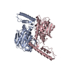
- Structure visualization
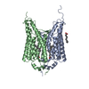
| Structure viewer | Molecule: MolmilJmol/JSmol |
|---|
- Downloads & links
-Download
| PDBx/mmCIF format | 5zih.cif.gz | 137.7 KB | Display | PDBx/mmCIF format |
|---|---|---|---|---|
| PDB format | pdb5zih.ent.gz | 106.6 KB | Display | PDB format |
| PDBx/mmJSON format | 5zih.json.gz | Tree view | PDBx/mmJSON format | |
| Others |  Other downloads Other downloads |
-Validation report
| Arichive directory | https://data.pdbj.org/pub/pdb/validation_reports/zi/5zihftp://data.pdbj.org/pub/pdb/validation_reports/zi/5zih | HTTPS FTP |
|---|
-Related structure data
| Related structure data |  3ug9S S: Starting model for refinement |
|---|---|
| Similar structure data | |
| Experimental dataset #1 | Data reference: 10.5281/zenodo.1319974 / Data set type: diffraction image data / Metadata reference: 10.5281/zenodo.1319974 |

-Links
 PDBj
PDBj


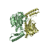
- Assembly
| Deposited unit | 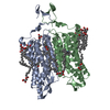
| ||||||||
|---|---|---|---|---|---|---|---|---|---|
| 1 |
| ||||||||
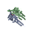
| Unit cell |
|
-Components
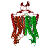
| #1: Protein | Mass: 38959.156 Da / Num. of mol.: 2 Source method: isolated from a genetically manipulated source Details: Chrimson with substituted N-terminal CrChR1 sequence Source: (gene. exp.) Chlamydomonas reinhardtii (plant), (gene. exp.) Chlamydomonas noctigama (plant)Gene: CSOA / Plasmid: pFastBac1 / Cell line (production host): Sf9 / Production host:   Spodoptera frugiperda (fall armyworm) / References: UniProt: Q8L435 Spodoptera frugiperda (fall armyworm) / References: UniProt: Q8L435#2: Chemical | ChemComp-OLC / (   Mass: 356.540 Da / Num. of mol.: 19 / Source method: obtained synthetically / Formula: C21H40O4 Mass: 356.540 Da / Num. of mol.: 19 / Source method: obtained synthetically / Formula: C21H40O4#3: Water | ChemComp-HOH / |  Mass: 18.015 Da / Num. of mol.: 37 / Source method: isolated from a natural source / Formula: H2O Mass: 18.015 Da / Num. of mol.: 37 / Source method: isolated from a natural source / Formula: H2O |
|---|
-Experimental details
-Experiment
| Experiment | Method: X-RAY DIFFRACTION / Number of used crystals: 1 |
|---|
- Sample preparation
| Crystal | Density Matthews: 2.73 Å3/Da / Density % sol: 54.95 % |
|---|---|
| Crystal grow | Temperature: 298 K / Method: lipidic cubic phase / pH: 7 / Details: PEG500DME, Na citrate, Na malonate, sarcosine |
-Data collection
| Diffraction | Mean temperature: 100 K |
|---|---|
| Diffraction source | Source: SYNCHROTRON / Site: SPring-8 / Beamline: BL32XU / Wavelength: 1 Å |
| Detector | Type: DECTRIS EIGER X 9M / Detector: PIXEL / Date: Jul 23, 2016 |
| Radiation | Protocol: SINGLE WAVELENGTH / Monochromatic (M) / Laue (L): M / Scattering type: x-ray |
| Radiation wavelength | Wavelength: 1 Å / Relative weight: 1 |
| Reflection | Resolution: 2.6→50 Å / Num. obs: 30180 / % possible obs: 100 % / Redundancy: 9.3 % / Biso Wilson estimate: 24.96 Å2 / CC1/2: 0.98 / Rrim(I) all: 0.144 / Χ2: 1.237 / Net I/σ(I): 13.8 / Num. measured all: 276798 / Scaling rejects: 214 |
| Reflection shell | Resolution: 2.6→2.69 Å / Redundancy: 9.2 % / Mean I/σ(I) obs: 1.7 / Num. measured obs: 41376 / Num. possible: 4768 / Num. unique obs: 4767 / CC1/2: 0.595 / Rrim(I) all: 0.591 / % possible all: 100 |
- Processing
| Software |
| ||||||||||||||||||||||||||||||||||||||||||||||||||||||||||||||||||||||||||||||||||||||||||||||||||||||||||||||||||||||||||||||||||||||||||||
|---|---|---|---|---|---|---|---|---|---|---|---|---|---|---|---|---|---|---|---|---|---|---|---|---|---|---|---|---|---|---|---|---|---|---|---|---|---|---|---|---|---|---|---|---|---|---|---|---|---|---|---|---|---|---|---|---|---|---|---|---|---|---|---|---|---|---|---|---|---|---|---|---|---|---|---|---|---|---|---|---|---|---|---|---|---|---|---|---|---|---|---|---|---|---|---|---|---|---|---|---|---|---|---|---|---|---|---|---|---|---|---|---|---|---|---|---|---|---|---|---|---|---|---|---|---|---|---|---|---|---|---|---|---|---|---|---|---|---|---|---|---|
| Refinement | Method to determine structure: MOLECULAR REPLACEMENT Starting model: 3UG9 Resolution: 2.6→48.818 Å / SU ML: 0.36 / Cross valid method: THROUGHOUT / σ(F): 1.34 / Phase error: 31.76 / Stereochemistry target values: ML
| ||||||||||||||||||||||||||||||||||||||||||||||||||||||||||||||||||||||||||||||||||||||||||||||||||||||||||||||||||||||||||||||||||||||||||||
| Solvent computation | Shrinkage radii: 0.9 Å / VDW probe radii: 1.11 Å / Solvent model: FLAT BULK SOLVENT MODEL | ||||||||||||||||||||||||||||||||||||||||||||||||||||||||||||||||||||||||||||||||||||||||||||||||||||||||||||||||||||||||||||||||||||||||||||
| Displacement parameters | Biso max: 150.21 Å2 / Biso mean: 38.6213 Å2 / Biso min: 3.56 Å2 | ||||||||||||||||||||||||||||||||||||||||||||||||||||||||||||||||||||||||||||||||||||||||||||||||||||||||||||||||||||||||||||||||||||||||||||
| Refinement step | Cycle: final / Resolution: 2.6→48.818 Å
| ||||||||||||||||||||||||||||||||||||||||||||||||||||||||||||||||||||||||||||||||||||||||||||||||||||||||||||||||||||||||||||||||||||||||||||
| LS refinement shell | Refine-ID: X-RAY DIFFRACTION / Rfactor Rfree error: 0 / Total num. of bins used: 19
|
