 Movie
Movie Controller

[English] 日本語
 Yorodumi
- PDB-5dg5: CRYSTAL STRUCTURE OF THE TYROSINE KINASE DOMAIN OF THE HEPATOCYTE... -
+ Open data

- Basic information
| Entry | Database: PDB / ID: 5dg5 | ||||||
|---|---|---|---|---|---|---|---|
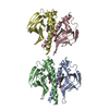
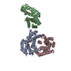
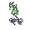
| Title | CRYSTAL STRUCTURE OF THE TYROSINE KINASE DOMAIN OF THE HEPATOCYTE GROWTH FACTOR RECEPTOR C-MET IN COMPLEX WITH ALTIRATINIB ANALOG DP-4157 | ||||||
 Components Components | Hepatocyte growth factor receptor | ||||||
 Keywords Keywords | TRANSFERASE/TRANSFERASE INHIBITOR / TYROSINE KINASE DOMAIN / HEPATOCYTE GROWTH FACTOR RECEPTOR C-MET / C-MET / ALTIRATINIB ANALOG / DP-4157 / TRANSFERASE-TRANSFERASE INHIBITOR complex | ||||||
| Function / homology |  Function and homology information Function and homology informationhepatocyte growth factor receptor activity / Drug-mediated inhibition of MET activation / MET activates STAT3 / negative regulation of hydrogen peroxide-mediated programmed cell death / MET Receptor Activation / MET interacts with TNS proteins / endothelial cell morphogenesis / semaphorin receptor activity / MET receptor recycling / pancreas development ...hepatocyte growth factor receptor activity / Drug-mediated inhibition of MET activation / MET activates STAT3 / negative regulation of hydrogen peroxide-mediated programmed cell death / MET Receptor Activation / MET interacts with TNS proteins / endothelial cell morphogenesis / semaphorin receptor activity / MET receptor recycling / pancreas development / MET activates PTPN11 / hepatocyte growth factor receptor signaling pathway / MET activates RAP1 and RAC1 / Sema4D mediated inhibition of cell attachment and migration / positive regulation of endothelial cell chemotaxis / MET activates PI3K/AKT signaling / MET activates PTK2 signaling / branching morphogenesis of an epithelial tube / positive chemotaxis / semaphorin-plexin signaling pathway / Regulation of MITF-M-dependent genes involved in cell cycle and proliferation / MET activates RAS signaling / MECP2 regulates neuronal receptors and channels / cell surface receptor protein tyrosine kinase signaling pathway / basal plasma membrane / molecular function activator activity / negative regulation of autophagy / InlB-mediated entry of Listeria monocytogenes into host cell / liver development / excitatory postsynaptic potential / receptor protein-tyrosine kinase / Negative regulation of MET activity / Constitutive Signaling by Aberrant PI3K in Cancer / neuron differentiation / PIP3 activates AKT signaling / PI5P, PP2A and IER3 Regulate PI3K/AKT Signaling / RAF/MAP kinase cascade / protein tyrosine kinase activity / protein phosphatase binding / cell surface receptor signaling pathway / signaling receptor complex / postsynapse / cell surface / positive regulation of transcription by RNA polymerase II / extracellular region / ATP binding / membrane / identical protein binding / plasma membrane Similarity search - Function | ||||||
| Biological species |  Homo sapiens (human) Homo sapiens (human) | ||||||
| Method |  X-RAY DIFFRACTION / SYNCHROTRON / MOLECULAR REPLACEMENT / Resolution: 2.6 Å X-RAY DIFFRACTION / SYNCHROTRON / MOLECULAR REPLACEMENT / Resolution: 2.6 Å | ||||||
 Authors Authors | Smith, B.D. / Kaufman, M.D. / Leary, C.B. / Turner, B.A. / Wise, S.A. / Ahn, Y.M. / Booth, R.J. / Caldwell, T.M. / Ensinger, C.L. / Hood, M.M. ...Smith, B.D. / Kaufman, M.D. / Leary, C.B. / Turner, B.A. / Wise, S.A. / Ahn, Y.M. / Booth, R.J. / Caldwell, T.M. / Ensinger, C.L. / Hood, M.M. / Lu, W.-P. / Patt, T.W. / Patt, W.C. / Rutkoski, T.J. / Samarakoon, T. / Telikepalli, H. / Vogeti, L. / Vogeti, S. / Yates, K.M. / Chun, L. / Stewart, L.J. / Clare, M. / Flynn, D.L. | ||||||
 Citation Citation | Journal: Mol.Cancer Ther. / Year: 2015 Title: Altiratinib Inhibits Tumor Growth, Invasion, Angiogenesis, and Microenvironment-Mediated Drug Resistance via Balanced Inhibition of MET, TIE2, and VEGFR2. Authors: Smith, B.D. / Kaufman, M.D. / Leary, C.B. / Turner, B.A. / Wise, S.C. / Ahn, Y.M. / Booth, R.J. / Caldwell, T.M. / Ensinger, C.L. / Hood, M.M. / Lu, W.P. / Patt, T.W. / Patt, W.C. / ...Authors: Smith, B.D. / Kaufman, M.D. / Leary, C.B. / Turner, B.A. / Wise, S.C. / Ahn, Y.M. / Booth, R.J. / Caldwell, T.M. / Ensinger, C.L. / Hood, M.M. / Lu, W.P. / Patt, T.W. / Patt, W.C. / Rutkoski, T.J. / Samarakoon, T. / Telikepalli, H. / Vogeti, L. / Vogeti, S. / Yates, K.M. / Chun, L. / Stewart, L.J. / Clare, M. / Flynn, D.L. | ||||||
| History |
|
- Structure visualization
| Structure viewer | Molecule: MolmilJmol/JSmol |
|---|
- Downloads & links
-Download
| PDBx/mmCIF format | 5dg5.cif.gz | 132 KB | Display | PDBx/mmCIF format |
|---|---|---|---|---|
| PDB format | pdb5dg5.ent.gz | 100.5 KB | Display | PDB format |
| PDBx/mmJSON format | 5dg5.json.gz | Tree view | PDBx/mmJSON format | |
| Others |  Other downloads Other downloads |
-Validation report
| Arichive directory | https://data.pdbj.org/pub/pdb/validation_reports/dg/5dg5ftp://data.pdbj.org/pub/pdb/validation_reports/dg/5dg5 | HTTPS FTP |
|---|
-Related structure data
| Related structure data | 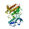 2g15S S: Starting model for refinement |
|---|---|
| Similar structure data |

-Links
 PDBj
PDBj


- Assembly
| Deposited unit | 
| ||||||||||||||||||
|---|---|---|---|---|---|---|---|---|---|---|---|---|---|---|---|---|---|---|---|
| 1 |
| ||||||||||||||||||
| Unit cell |
| ||||||||||||||||||
| Noncrystallographic symmetry (NCS) | NCS domain:
NCS domain segments: Component-ID: _ / Ens-ID: 1 / Beg auth comp-ID: THR / Beg label comp-ID: THR / End auth comp-ID: GLY / End label comp-ID: GLY / Refine code: _ / Auth seq-ID: 1050 - 1346 / Label seq-ID: 22 - 318
|
-Components
| #1: Protein | Mass: 35990.633 Da / Num. of mol.: 2 Source method: isolated from a genetically manipulated source Source: (gene. exp.) Homo sapiens (human) / Gene: MET / Production host:  References: UniProt: P08581, receptor protein-tyrosine kinase #2: Chemical |   Mass: 507.464 Da / Num. of mol.: 2 Mass: 507.464 Da / Num. of mol.: 2Source method: isolated from a genetically manipulated source Formula: C26H20F3N5O3 #3: Water | ChemComp-HOH / |  Mass: 18.015 Da / Num. of mol.: 33 / Source method: isolated from a natural source / Formula: H2O Mass: 18.015 Da / Num. of mol.: 33 / Source method: isolated from a natural source / Formula: H2O |
|---|
-Experimental details
-Experiment
| Experiment | Method: X-RAY DIFFRACTION |
|---|
- Sample preparation
| Crystal | Density Matthews: 2.69 Å3/Da / Density % sol: 54 % |
|---|---|
| Crystal grow | Temperature: 290 K / Method: vapor diffusion, sitting drop Details: Protein at 9.5mg/ml in 20 mM Tris pH 8.5, 100mM NaCl, 14mM 2-mercaptoethanol with 5-molar excess of compound; crystallization condition: 1.0M diammonium hydrogen phosphate, 0.2M sodium ...Details: Protein at 9.5mg/ml in 20 mM Tris pH 8.5, 100mM NaCl, 14mM 2-mercaptoethanol with 5-molar excess of compound; crystallization condition: 1.0M diammonium hydrogen phosphate, 0.2M sodium chloride, 0.1M citrate pH 5.0 and 7.5% glycerol |
-Data collection
| Diffraction | Mean temperature: 100 K | |||||||||||||||||||||||||||||||||||||||||||||||||||||||||||||||||||||||||||||||||||||||||||||||||||
|---|---|---|---|---|---|---|---|---|---|---|---|---|---|---|---|---|---|---|---|---|---|---|---|---|---|---|---|---|---|---|---|---|---|---|---|---|---|---|---|---|---|---|---|---|---|---|---|---|---|---|---|---|---|---|---|---|---|---|---|---|---|---|---|---|---|---|---|---|---|---|---|---|---|---|---|---|---|---|---|---|---|---|---|---|---|---|---|---|---|---|---|---|---|---|---|---|---|---|---|---|
| Diffraction source | Source: SYNCHROTRON / Site: ALS  / Beamline: 5.0.2 / Wavelength: 0.9999 Å / Beamline: 5.0.2 / Wavelength: 0.9999 Å | |||||||||||||||||||||||||||||||||||||||||||||||||||||||||||||||||||||||||||||||||||||||||||||||||||
| Detector | Type: ADSC QUANTUM 315r / Detector: CCD / Date: Apr 17, 2009 | |||||||||||||||||||||||||||||||||||||||||||||||||||||||||||||||||||||||||||||||||||||||||||||||||||
| Radiation | Monochromator: Si(111) / Protocol: SINGLE WAVELENGTH / Monochromatic (M) / Laue (L): M / Scattering type: x-ray | |||||||||||||||||||||||||||||||||||||||||||||||||||||||||||||||||||||||||||||||||||||||||||||||||||
| Radiation wavelength | Wavelength: 0.9999 Å / Relative weight: 1 | |||||||||||||||||||||||||||||||||||||||||||||||||||||||||||||||||||||||||||||||||||||||||||||||||||
| Reflection | Resolution: 2.6→20 Å / Num. obs: 23076 / % possible obs: 98.3 % / Observed criterion σ(I): -3 / Redundancy: 3.2 % / Biso Wilson estimate: 47.861 Å2 / Rmerge F obs: 0.211 / Rmerge(I) obs: 0.1 / Rrim(I) all: 0.12 / Χ2: 0.96 / Net I/σ(I): 11.36 / Num. measured all: 73855 | |||||||||||||||||||||||||||||||||||||||||||||||||||||||||||||||||||||||||||||||||||||||||||||||||||
| Reflection shell | Diffraction-ID: 1 / Rejects: _
|
- Processing
| Software |
| |||||||||||||||||||||||||||||||||||||||||||||||||||||||||||||||||||||||||||
|---|---|---|---|---|---|---|---|---|---|---|---|---|---|---|---|---|---|---|---|---|---|---|---|---|---|---|---|---|---|---|---|---|---|---|---|---|---|---|---|---|---|---|---|---|---|---|---|---|---|---|---|---|---|---|---|---|---|---|---|---|---|---|---|---|---|---|---|---|---|---|---|---|---|---|---|---|
| Refinement | Method to determine structure: MOLECULAR REPLACEMENT Starting model: PDB entry 2g15 Resolution: 2.6→19.65 Å / Cor.coef. Fo:Fc: 0.941 / Cor.coef. Fo:Fc free: 0.917 / WRfactor Rfree: 0.2096 / WRfactor Rwork: 0.1759 / FOM work R set: 0.755 / SU B: 14.157 / SU ML: 0.283 / SU R Cruickshank DPI: 0.6743 / SU Rfree: 0.3023 / Cross valid method: THROUGHOUT / σ(F): 0 / ESU R: 0.674 / ESU R Free: 0.302 / Stereochemistry target values: MAXIMUM LIKELIHOOD Details: HYDROGENS HAVE BEEN ADDED IN THE RIDING POSITIONS U VALUES : REFINED INDIVIDUALLY
| |||||||||||||||||||||||||||||||||||||||||||||||||||||||||||||||||||||||||||
| Solvent computation | Ion probe radii: 0.8 Å / Shrinkage radii: 0.8 Å / VDW probe radii: 1.2 Å / Solvent model: MASK | |||||||||||||||||||||||||||||||||||||||||||||||||||||||||||||||||||||||||||
| Displacement parameters | Biso max: 103.15 Å2 / Biso mean: 43.412 Å2 / Biso min: 15.89 Å2
| |||||||||||||||||||||||||||||||||||||||||||||||||||||||||||||||||||||||||||
| Refinement step | Cycle: final / Resolution: 2.6→19.65 Å
| |||||||||||||||||||||||||||||||||||||||||||||||||||||||||||||||||||||||||||
| Refine LS restraints |
| |||||||||||||||||||||||||||||||||||||||||||||||||||||||||||||||||||||||||||
| Refine LS restraints NCS | Ens-ID: 1 / Number: 17561 / Refine-ID: X-RAY DIFFRACTION / Type: interatomic distance / Rms dev position: 0.08 Å / Weight position: 0.05
| |||||||||||||||||||||||||||||||||||||||||||||||||||||||||||||||||||||||||||
| LS refinement shell | Resolution: 2.6→2.666 Å / Total num. of bins used: 20
|
