 Movie
Movie Controller

[English] 日本語
 Yorodumi
- PDB-5dcq: Crystal structure of bacterial adhesin, FNE from Streptococcus eq... -
+ Open data

- Basic information
| Entry | Database: PDB / ID: 5dcq | ||||||
|---|---|---|---|---|---|---|---|
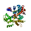
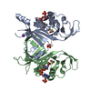
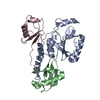
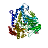
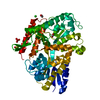
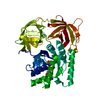
| Title | Crystal structure of bacterial adhesin, FNE from Streptococcus equi spp. equi. | ||||||
 Components Components |
| ||||||
 Keywords Keywords | STRUCTURAL PROTEIN / adhesin / artificial repeat proteins / complex / extracellular matrix / pilus / thioester bond | ||||||
| Function / homology | Uncharacterised domain CHP03934, TQXA / Thioester domain / Thioester domain / FORMIC ACID / Fibronectin-binding protein Function and homology information Function and homology information | ||||||
| Biological species | synthetic construct (others) Streptococcus equi subsp. equi (bacteria) Streptococcus equi subsp. equi (bacteria) | ||||||
| Method |  X-RAY DIFFRACTION / SYNCHROTRON / MOLECULAR REPLACEMENT / Resolution: 1.83 Å X-RAY DIFFRACTION / SYNCHROTRON / MOLECULAR REPLACEMENT / Resolution: 1.83 Å | ||||||
 Authors Authors | Tiouajni, M. / Graille, M. / van Tilbeurgh, H. | ||||||
 Citation Citation | Journal: FEBS J. / Year: 2014 Title: Structural and functional analysis of the fibronectin-binding protein FNE from Streptococcus equi spp. equi. Authors: Tiouajni, M. / Durand, D. / Blondeau, K. / Graille, M. / Urvoas, A. / Valerio-Lepiniec, M. / Guellouz, A. / Aumont-Nicaise, M. / Minard, P. / van Tilbeurgh, H. | ||||||
| History |
|
- Structure visualization
| Structure viewer | Molecule: MolmilJmol/JSmol |
|---|
- Downloads & links
-Download
| PDBx/mmCIF format | 5dcq.cif.gz | 459.8 KB | Display | PDBx/mmCIF format |
|---|---|---|---|---|
| PDB format | pdb5dcq.ent.gz | 377.6 KB | Display | PDB format |
| PDBx/mmJSON format | 5dcq.json.gz | Tree view | PDBx/mmJSON format | |
| Others |  Other downloads Other downloads |
-Validation report
| Arichive directory | https://data.pdbj.org/pub/pdb/validation_reports/dc/5dcqftp://data.pdbj.org/pub/pdb/validation_reports/dc/5dcq | HTTPS FTP |
|---|
-Related structure data
| Related structure data |  2xi9S  3ltjS  4pfg S: Starting model for refinement |
|---|---|
| Similar structure data |


-Links
 PDBj
PDBj- Assembly
| Deposited unit | 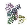
| ||||||||
|---|---|---|---|---|---|---|---|---|---|
| 1 | 
| ||||||||
| 2 | 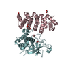
| ||||||||
| 3 | 
| ||||||||
| 4 | 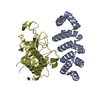
| ||||||||
| 5 | 
| ||||||||
| Unit cell |
|
-Components
| #1: Protein | Mass: 18678.240 Da / Num. of mol.: 3 Source method: isolated from a genetically manipulated source Source: (gene. exp.) synthetic construct (others) / Production host: #2: Protein | Mass: 30697.811 Da / Num. of mol.: 3 / Fragment: UNP residues 35-299 Source method: isolated from a genetically manipulated source Source: (gene. exp.) Streptococcus equi subsp. equi (bacteria)Gene: fne Production host: References: UniProt: Q93ED6 #3: Chemical | ChemComp-FMT /   Mass: 46.025 Da / Num. of mol.: 12 / Source method: obtained synthetically / Formula: CH2O2 Mass: 46.025 Da / Num. of mol.: 12 / Source method: obtained synthetically / Formula: CH2O2#4: Water | ChemComp-HOH / |  Mass: 18.015 Da / Num. of mol.: 1102 / Source method: isolated from a natural source / Formula: H2O Mass: 18.015 Da / Num. of mol.: 1102 / Source method: isolated from a natural source / Formula: H2O |
|---|
-Experimental details
-Experiment
| Experiment | Method: X-RAY DIFFRACTION / Number of used crystals: 1 |
|---|
- Sample preparation
| Crystal | Density Matthews: 2.34 Å3/Da / Density % sol: 47.5 % |
|---|---|
| Crystal grow | Temperature: 277.15 K / Method: vapor diffusion, sitting drop / Details: magnesium formate, PEG 3350 |
-Data collection
| Diffraction | Mean temperature: 153 K |
|---|---|
| Diffraction source | Source: SYNCHROTRON / Site: SOLEIL  / Beamline: PROXIMA 1 / Wavelength: 0.972 Å / Beamline: PROXIMA 1 / Wavelength: 0.972 Å |
| Detector | Type: DECTRIS PILATUS 6M / Detector: PIXEL / Date: Feb 23, 2012 |
| Radiation | Protocol: SINGLE WAVELENGTH / Monochromatic (M) / Laue (L): M / Scattering type: x-ray |
| Radiation wavelength | Wavelength: 0.972 Å / Relative weight: 1 |
| Reflection | Resolution: 1.83→50 Å / Num. obs: 91861 / % possible obs: 97.78 % / Redundancy: 2.3 % / Rsym value: 0.06 / Net I/σ(I): 14.2 |
| Reflection shell | Resolution: 1.83→1.94 Å / Redundancy: 2.3 % / Mean I/σ(I) obs: 1.9 / Rsym value: 0.642 / % possible all: 97.7 |
- Processing
| Software |
| |||||||||||||||||||||||||||||||||||||||||||||||||||||||||||||||||||||||||||||||||||||||||||||||||||||||||||||||||||||||||||||||||||||||||||||||||||||||||||||||||||||||||||||||
|---|---|---|---|---|---|---|---|---|---|---|---|---|---|---|---|---|---|---|---|---|---|---|---|---|---|---|---|---|---|---|---|---|---|---|---|---|---|---|---|---|---|---|---|---|---|---|---|---|---|---|---|---|---|---|---|---|---|---|---|---|---|---|---|---|---|---|---|---|---|---|---|---|---|---|---|---|---|---|---|---|---|---|---|---|---|---|---|---|---|---|---|---|---|---|---|---|---|---|---|---|---|---|---|---|---|---|---|---|---|---|---|---|---|---|---|---|---|---|---|---|---|---|---|---|---|---|---|---|---|---|---|---|---|---|---|---|---|---|---|---|---|---|---|---|---|---|---|---|---|---|---|---|---|---|---|---|---|---|---|---|---|---|---|---|---|---|---|---|---|---|---|---|---|---|---|---|
| Refinement | Method to determine structure: MOLECULAR REPLACEMENT Starting model: 2XI9, 3LTJ Resolution: 1.83→50 Å / Cor.coef. Fo:Fc: 0.967 / Cor.coef. Fo:Fc free: 0.948 / SU B: 6.309 / SU ML: 0.094 / Cross valid method: THROUGHOUT / σ(F): 0 / ESU R: 0.128 / ESU R Free: 0.12 / Stereochemistry target values: MAXIMUM LIKELIHOOD Details: HYDROGENS HAVE BEEN ADDED IN THE RIDING POSITIONS U VALUES : RESIDUAL ONLY
| |||||||||||||||||||||||||||||||||||||||||||||||||||||||||||||||||||||||||||||||||||||||||||||||||||||||||||||||||||||||||||||||||||||||||||||||||||||||||||||||||||||||||||||||
| Solvent computation | Ion probe radii: 0.8 Å / Shrinkage radii: 0.8 Å / VDW probe radii: 1.2 Å / Solvent model: MASK | |||||||||||||||||||||||||||||||||||||||||||||||||||||||||||||||||||||||||||||||||||||||||||||||||||||||||||||||||||||||||||||||||||||||||||||||||||||||||||||||||||||||||||||||
| Displacement parameters | Biso max: 93.72 Å2 / Biso mean: 19.445 Å2 / Biso min: 7.14 Å2
| |||||||||||||||||||||||||||||||||||||||||||||||||||||||||||||||||||||||||||||||||||||||||||||||||||||||||||||||||||||||||||||||||||||||||||||||||||||||||||||||||||||||||||||||
| Refinement step | Cycle: final / Resolution: 1.83→50 Å
| |||||||||||||||||||||||||||||||||||||||||||||||||||||||||||||||||||||||||||||||||||||||||||||||||||||||||||||||||||||||||||||||||||||||||||||||||||||||||||||||||||||||||||||||
| Refine LS restraints |
| |||||||||||||||||||||||||||||||||||||||||||||||||||||||||||||||||||||||||||||||||||||||||||||||||||||||||||||||||||||||||||||||||||||||||||||||||||||||||||||||||||||||||||||||
| LS refinement shell | Resolution: 1.826→1.873 Å / Total num. of bins used: 20
| |||||||||||||||||||||||||||||||||||||||||||||||||||||||||||||||||||||||||||||||||||||||||||||||||||||||||||||||||||||||||||||||||||||||||||||||||||||||||||||||||||||||||||||||
| Refinement TLS params. | Method: refined / Refine-ID: X-RAY DIFFRACTION
| |||||||||||||||||||||||||||||||||||||||||||||||||||||||||||||||||||||||||||||||||||||||||||||||||||||||||||||||||||||||||||||||||||||||||||||||||||||||||||||||||||||||||||||||
| Refinement TLS group |
|
