 Movie
Movie Controller

[English] 日本語
 Yorodumi
- PDB-1gjv: Branched-chain alpha-ketoacid dehydrogenase kinase (BCK) complxed... -
+ Open data

- Basic information
| Entry | Database: PDB / ID: 1gjv | ||||||
|---|---|---|---|---|---|---|---|
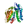
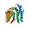
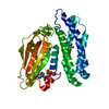
| Title | Branched-chain alpha-ketoacid dehydrogenase kinase (BCK) complxed with ATP-gamma-S | ||||||
 Components Components | [3-METHYL-2-OXOBUTANOATE DEHYDROGENASE [LIPOAMIDE]] KINASE | ||||||
 Keywords Keywords | TRANSFERASE / MITOCHONDRIAL PROTEIN KINASE / POTASSIUM | ||||||
| Function / homology |  Function and homology information Function and homology information[3-methyl-2-oxobutanoate dehydrogenase (acetyl-transferring)] kinase / [3-methyl-2-oxobutanoate dehydrogenase (acetyl-transferring)] kinase activity / Branched-chain amino acid catabolism / pyruvate dehydrogenase (acetyl-transferring) kinase activity / L-valine catabolic process / regulation of pyruvate decarboxylation to acetyl-CoA / L-isoleucine catabolic process / L-leucine catabolic process / oxoglutarate dehydrogenase complex / branched-chain amino acid catabolic process ...[3-methyl-2-oxobutanoate dehydrogenase (acetyl-transferring)] kinase / [3-methyl-2-oxobutanoate dehydrogenase (acetyl-transferring)] kinase activity / Branched-chain amino acid catabolism / pyruvate dehydrogenase (acetyl-transferring) kinase activity / L-valine catabolic process / regulation of pyruvate decarboxylation to acetyl-CoA / L-isoleucine catabolic process / L-leucine catabolic process / oxoglutarate dehydrogenase complex / branched-chain amino acid catabolic process / lipid biosynthetic process / protein serine/threonine phosphatase activity / regulation of glucose metabolic process / spermatogenesis / protein kinase activity / non-specific serine/threonine protein kinase / mitochondrial matrix / protein serine kinase activity / protein serine/threonine kinase activity / mitochondrion / ATP binding Similarity search - Function | ||||||
| Biological species |  | ||||||
| Method |  X-RAY DIFFRACTION / MOLECULAR REPLACEMENT / Resolution: 2.7 Å X-RAY DIFFRACTION / MOLECULAR REPLACEMENT / Resolution: 2.7 Å | ||||||
 Authors Authors | Machius, M. / Chuang, J.L. / Wynn, M.R. / Tomchick, D.R. / Chuang, D.T. | ||||||
 Citation Citation | Journal: Proc.Natl.Acad.Sci.USA / Year: 2001 Title: Structure of Rat Bckd Kinase: Nucleotide-Induced Domain Communication in a Mitochondrial Protein Kinase. Authors: Machius, M. / Chuang, J.L. / Wynn, M.R. / Tomchick, D.R. / Chuang, D.T. | ||||||
| History |
|
- Structure visualization
| Structure viewer | Molecule: MolmilJmol/JSmol |
|---|
- Downloads & links
-Download
| PDBx/mmCIF format | 1gjv.cif.gz | 81.3 KB | Display | PDBx/mmCIF format |
|---|---|---|---|---|
| PDB format | pdb1gjv.ent.gz | 60 KB | Display | PDB format |
| PDBx/mmJSON format | 1gjv.json.gz | Tree view | PDBx/mmJSON format | |
| Others |  Other downloads Other downloads |
-Validation report
| Arichive directory | https://data.pdbj.org/pub/pdb/validation_reports/gj/1gjvftp://data.pdbj.org/pub/pdb/validation_reports/gj/1gjv | HTTPS FTP |
|---|
-Related structure data


-Links
 PDBj
PDBj


- Assembly
| Deposited unit | 
| ||||||||
|---|---|---|---|---|---|---|---|---|---|
| 1 |
| ||||||||
| Unit cell |
|
-Components
-Protein , 1 types, 1 molecules A
| #1: Protein | [ Mass: 44261.582 Da / Num. of mol.: 1 Source method: isolated from a genetically manipulated source Details: C-TERMINAL HIS6-TAG / Source: (gene. exp.)  |
|---|
-Non-polymers , 5 types, 78 molecules 


| #2: Chemical | ChemComp-CL /  Mass: 35.453 Da / Num. of mol.: 5 / Source method: obtained synthetically / Formula: Cl Mass: 35.453 Da / Num. of mol.: 5 / Source method: obtained synthetically / Formula: Cl#3: Chemical | ChemComp-MG / |  Mass: 24.305 Da / Num. of mol.: 1 / Source method: obtained synthetically / Formula: Mg Mass: 24.305 Da / Num. of mol.: 1 / Source method: obtained synthetically / Formula: Mg#4: Chemical | ChemComp-K / |  Mass: 39.098 Da / Num. of mol.: 1 / Source method: obtained synthetically / Formula: K Mass: 39.098 Da / Num. of mol.: 1 / Source method: obtained synthetically / Formula: K#5: Chemical | ChemComp-AGS / |  Mass: 523.247 Da / Num. of mol.: 1 / Source method: obtained synthetically / Formula: C10H16N5O12P3S / Comment: ATP-gamma-S, energy-carrying molecule analogue*YM Mass: 523.247 Da / Num. of mol.: 1 / Source method: obtained synthetically / Formula: C10H16N5O12P3S / Comment: ATP-gamma-S, energy-carrying molecule analogue*YM#6: Water | ChemComp-HOH / | Mass: 18.015 Da / Num. of mol.: 70 / Source method: isolated from a natural source / Formula: H2O |
|---|
-Details
| Sequence details | CRYSTALLIZ |
|---|
-Experimental details
-Experiment
| Experiment | Method: X-RAY DIFFRACTION / Number of used crystals: 1 |
|---|
- Sample preparation
| Crystal | Density Matthews: 3.4 Å3/Da / Density % sol: 64 % | ||||||||||||||||||||||||||||||||||||||||||||||||||||||||||||||||||||||||||||||||||||||||||||||||
|---|---|---|---|---|---|---|---|---|---|---|---|---|---|---|---|---|---|---|---|---|---|---|---|---|---|---|---|---|---|---|---|---|---|---|---|---|---|---|---|---|---|---|---|---|---|---|---|---|---|---|---|---|---|---|---|---|---|---|---|---|---|---|---|---|---|---|---|---|---|---|---|---|---|---|---|---|---|---|---|---|---|---|---|---|---|---|---|---|---|---|---|---|---|---|---|---|---|
| Crystal grow | Temperature: 293 K / Method: vapor diffusion / pH: 7.5 Details: VAPOR DIFFUSION 20C EQUAL AMOUNTS OF BCK (20 MG/ML IN 50 MM HEPES PH 7.5, 1 M SODIUM CHLORIDE,250 MM POTASSIUM CHLORIDE, 300 MM ARGININE, 20 MM BETA-MERCAPTOETHANOL 2 MM BENZAMIDINE, 2 MM MG- ...Details: VAPOR DIFFUSION 20C EQUAL AMOUNTS OF BCK (20 MG/ML IN 50 MM HEPES PH 7.5, 1 M SODIUM CHLORIDE,250 MM POTASSIUM CHLORIDE, 300 MM ARGININE, 20 MM BETA-MERCAPTOETHANOL 2 MM BENZAMIDINE, 2 MM MG-ATPGAMMAS, 2 MM MAGNESIUM CHLORIDE, 0.5 MM PMSF, 10% (W/V) GLYCEROL) AND RESERVOIR (8%(W/V) PEG-6000, 5% (V/V) ETHYLENE GLYCOL, 1 M NACL, 20 MM BETA-MERCAPTOETHANOL) | ||||||||||||||||||||||||||||||||||||||||||||||||||||||||||||||||||||||||||||||||||||||||||||||||
| Crystal grow | *PLUS Temperature: 20 ℃ / Method: vapor diffusion | ||||||||||||||||||||||||||||||||||||||||||||||||||||||||||||||||||||||||||||||||||||||||||||||||
| Components of the solutions | *PLUS
|
-Data collection
| Diffraction | Mean temperature: 100 K |
|---|---|
| Diffraction source | Source: ROTATING ANODE / Type: RIGAKU RU200 / Wavelength: 1.5418 |
| Detector | Type: MSC / Detector: IMAGE PLATE / Date: Apr 15, 2001 / Details: OSMIC MIRROR SYSTEM |
| Radiation | Monochromator: OSMIC MIRROR SYSTEM / Protocol: SINGLE WAVELENGTH / Monochromatic (M) / Laue (L): M / Scattering type: x-ray |
| Radiation wavelength | Wavelength: 1.5418 Å / Relative weight: 1 |
| Reflection | Resolution: 2.7→36.9 Å / Num. obs: 17401 / % possible obs: 99.6 % / Observed criterion σ(I): -3 / Redundancy: 4.6 % / Biso Wilson estimate: 70 Å2 / Rmerge(I) obs: 0.053 / Net I/σ(I): 24.4 |
| Reflection shell | Resolution: 2.7→2.8 Å / Redundancy: 3.6 % / Rmerge(I) obs: 0.533 / Mean I/σ(I) obs: 2 / % possible all: 96.8 |
| Reflection | *PLUS Rmerge(I) obs: 0.061 |
| Reflection shell | *PLUS Highest resolution: 2.7 Å / Lowest resolution: 2.8 Å / % possible obs: 96.8 % / Rmerge(I) obs: 0.582 |
- Processing
| Software |
| ||||||||||||||||||||||||||||||||||||||||||||||||||||||||||||||||||||||||||||||||
|---|---|---|---|---|---|---|---|---|---|---|---|---|---|---|---|---|---|---|---|---|---|---|---|---|---|---|---|---|---|---|---|---|---|---|---|---|---|---|---|---|---|---|---|---|---|---|---|---|---|---|---|---|---|---|---|---|---|---|---|---|---|---|---|---|---|---|---|---|---|---|---|---|---|---|---|---|---|---|---|---|---|
| Refinement | Method to determine structure: MOLECULAR REPLACEMENT Starting model: BRANCHED-CHAIN ALPHA-KETOACID DEHYDROGENASE KINASE (BCK) SELENOMETHIONINE VARIANT Resolution: 2.7→36.94 Å / Rfactor Rfree error: 0.007 / Data cutoff high absF: 298048.8 / Isotropic thermal model: RESTRAINED / Cross valid method: THROUGHOUT / σ(F): 0 Details: REGIONS: 1-37, 307-335, 379-388 WERE NOT SEEN IN THE REFINEMENT
| ||||||||||||||||||||||||||||||||||||||||||||||||||||||||||||||||||||||||||||||||
| Solvent computation | Solvent model: FLAT MODEL / Bsol: 51.4908 Å2 / ksol: 0.354628 e/Å3 | ||||||||||||||||||||||||||||||||||||||||||||||||||||||||||||||||||||||||||||||||
| Displacement parameters | Biso mean: 56.6 Å2
| ||||||||||||||||||||||||||||||||||||||||||||||||||||||||||||||||||||||||||||||||
| Refine analyze |
| ||||||||||||||||||||||||||||||||||||||||||||||||||||||||||||||||||||||||||||||||
| Refinement step | Cycle: LAST / Resolution: 2.7→36.94 Å
| ||||||||||||||||||||||||||||||||||||||||||||||||||||||||||||||||||||||||||||||||
| Refine LS restraints |
| ||||||||||||||||||||||||||||||||||||||||||||||||||||||||||||||||||||||||||||||||
| LS refinement shell | Resolution: 2.7→2.87 Å / Rfactor Rfree error: 0.026 / Total num. of bins used: 6
| ||||||||||||||||||||||||||||||||||||||||||||||||||||||||||||||||||||||||||||||||
| Xplor file |
| ||||||||||||||||||||||||||||||||||||||||||||||||||||||||||||||||||||||||||||||||
| Software | *PLUS Name: CNS / Version: 1 / Classification: refinement | ||||||||||||||||||||||||||||||||||||||||||||||||||||||||||||||||||||||||||||||||
| Refinement | *PLUS Rfactor obs: 0.231 / Rfactor Rwork: 0.281 | ||||||||||||||||||||||||||||||||||||||||||||||||||||||||||||||||||||||||||||||||
| Solvent computation | *PLUS | ||||||||||||||||||||||||||||||||||||||||||||||||||||||||||||||||||||||||||||||||
| Displacement parameters | *PLUS | ||||||||||||||||||||||||||||||||||||||||||||||||||||||||||||||||||||||||||||||||
| Refine LS restraints | *PLUS
|
