 Movie
Movie Controller

[English] 日本語
 Yorodumi
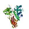
- PDB-2jri: Solution structure of the Josephin domain of Ataxin-3 in complex ... -
+ Open data

- Basic information
| Entry | Database: PDB / ID: 2jri | ||||||
|---|---|---|---|---|---|---|---|
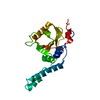
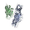
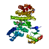
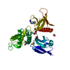
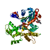
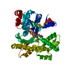
| Title | Solution structure of the Josephin domain of Ataxin-3 in complex with ubiquitin molecule. | ||||||
 Components Components |
| ||||||
 Keywords Keywords | HYDROLASE/SIGNALING PROTEIN / di-ubiquitin / Lys48-linked / Josephin domain of ataxin-3 / spinocerebellar ataxia type 3 protein / Alternative splicing / Hydrolase / Neurodegeneration / Nucleus / Phosphorylation / Polymorphism / Transcription / Transcription regulation / Triplet repeat expansion / HYDROLASE-SIGNALING PROTEIN COMPLEX | ||||||
| Function / homology |  Function and homology information Function and homology informationprotein localization to cytosolic proteasome complex / regulation of cell-substrate adhesion / positive regulation of ERAD pathway / monoubiquitinated protein deubiquitination / intermediate filament cytoskeleton organization / nuclear inclusion body / protein K48-linked deubiquitination / K48-linked deubiquitinase activity / cellular response to misfolded protein / positive regulation of ubiquitin-dependent protein catabolic process ...protein localization to cytosolic proteasome complex / regulation of cell-substrate adhesion / positive regulation of ERAD pathway / monoubiquitinated protein deubiquitination / intermediate filament cytoskeleton organization / nuclear inclusion body / protein K48-linked deubiquitination / K48-linked deubiquitinase activity / cellular response to misfolded protein / positive regulation of ubiquitin-dependent protein catabolic process / protein K63-linked deubiquitination / K63-linked deubiquitinase activity / exploration behavior / protein quality control for misfolded or incompletely synthesized proteins / FOXO-mediated transcription of oxidative stress, metabolic and neuronal genes / protein deubiquitination / negative regulation of TORC1 signaling / Maturation of protein E / Maturation of protein E / ER Quality Control Compartment (ERQC) / Myoclonic epilepsy of Lafora / FLT3 signaling by CBL mutants / IRAK2 mediated activation of TAK1 complex / Alpha-protein kinase 1 signaling pathway / Glycogen synthesis / IRAK1 recruits IKK complex / IRAK1 recruits IKK complex upon TLR7/8 or 9 stimulation / Prevention of phagosomal-lysosomal fusion / Endosomal Sorting Complex Required For Transport (ESCRT) / Membrane binding and targetting of GAG proteins / Regulation of TBK1, IKKε (IKBKE)-mediated activation of IRF3, IRF7 / Negative regulation of FLT3 / PTK6 Regulates RTKs and Their Effectors AKT1 and DOK1 / Regulation of TBK1, IKKε-mediated activation of IRF3, IRF7 upon TLR3 ligation / IRAK2 mediated activation of TAK1 complex upon TLR7/8 or 9 stimulation / Constitutive Signaling by NOTCH1 HD Domain Mutants / NOTCH2 Activation and Transmission of Signal to the Nucleus / TICAM1,TRAF6-dependent induction of TAK1 complex / TICAM1-dependent activation of IRF3/IRF7 / APC/C:Cdc20 mediated degradation of Cyclin B / Downregulation of ERBB4 signaling / APC-Cdc20 mediated degradation of Nek2A / Regulation of FZD by ubiquitination / p75NTR recruits signalling complexes / InlA-mediated entry of Listeria monocytogenes into host cells / TRAF6 mediated IRF7 activation in TLR7/8 or 9 signaling / NF-kB is activated and signals survival / TRAF6-mediated induction of TAK1 complex within TLR4 complex / Regulation of pyruvate metabolism / Pexophagy / Downregulation of ERBB2:ERBB3 signaling / cellular response to amino acid starvation / Regulation of innate immune responses to cytosolic DNA / NRIF signals cell death from the nucleus / Regulation of PTEN localization / VLDLR internalisation and degradation / Activated NOTCH1 Transmits Signal to the Nucleus / Synthesis of active ubiquitin: roles of E1 and E2 enzymes / Translesion synthesis by REV1 / TICAM1, RIP1-mediated IKK complex recruitment / Regulation of BACH1 activity / Translesion synthesis by POLK / JNK (c-Jun kinases) phosphorylation and activation mediated by activated human TAK1 / InlB-mediated entry of Listeria monocytogenes into host cell / MAP3K8 (TPL2)-dependent MAPK1/3 activation / Activation of IRF3, IRF7 mediated by TBK1, IKKε (IKBKE) / Downregulation of TGF-beta receptor signaling / Translesion synthesis by POLI / Josephin domain DUBs / Gap-filling DNA repair synthesis and ligation in GG-NER / IKK complex recruitment mediated by RIP1 / PINK1-PRKN Mediated Mitophagy / TGF-beta receptor signaling in EMT (epithelial to mesenchymal transition) / TNFR1-induced NF-kappa-B signaling pathway / Regulation of activated PAK-2p34 by proteasome mediated degradation / TCF dependent signaling in response to WNT / nucleotide-excision repair / Regulation of NF-kappa B signaling / activated TAK1 mediates p38 MAPK activation / Autodegradation of Cdh1 by Cdh1:APC/C / APC/C:Cdc20 mediated degradation of Securin / NOTCH3 Activation and Transmission of Signal to the Nucleus / Regulation of signaling by CBL / Negative regulators of DDX58/IFIH1 signaling / N-glycan trimming in the ER and Calnexin/Calreticulin cycle / Asymmetric localization of PCP proteins / Fanconi Anemia Pathway / Negative regulation of FGFR3 signaling / Ubiquitin-dependent degradation of Cyclin D / Peroxisomal protein import / Deactivation of the beta-catenin transactivating complex / SCF-beta-TrCP mediated degradation of Emi1 / NIK-->noncanonical NF-kB signaling / Stabilization of p53 / AUF1 (hnRNP D0) binds and destabilizes mRNA / TNFR2 non-canonical NF-kB pathway / Negative regulation of FGFR2 signaling / Negative regulation of FGFR4 signaling / Downregulation of SMAD2/3:SMAD4 transcriptional activity / Negative regulation of FGFR1 signaling Similarity search - Function | ||||||
| Biological species |  Homo sapiens (human) Homo sapiens (human) | ||||||
| Method | SOLUTION NMR / torsion angle dynamics | ||||||
| Model details | trascription/protein binding, machado-joseph disease protein, ubiquitin-proteasome pathway | ||||||
 Authors Authors | Nicastro, G. / Masino, L. / Esposito, V. / Menon, R. / Pastore, A. | ||||||
 Citation Citation | Journal: To be Published Title: Understanding the plasticity of the ubiquitin-protein recognition code: the josephin domain of ataxin-3 is a diubiquitin binding motif Authors: Nicastro, G. / Menon, R. / Masino, L. / Pastore, A. #1: Journal: Proc.Natl.Acad.Sci.USA / Year: 2005Title: The solution structure of the Josephin domain of ataxin-3: structural determinants for molecular recognition. Authors: Nicastro, G. / Masino, L. / Esposito, V. / Menon, R. / Pastore, A. | ||||||
| History |
|
- Structure visualization
| Structure viewer | Molecule: MolmilJmol/JSmol |
|---|
- Downloads & links
-Download
| PDBx/mmCIF format | 2jri.cif.gz | 2.1 MB | Display | PDBx/mmCIF format |
|---|---|---|---|---|
| PDB format | pdb2jri.ent.gz | 1.8 MB | Display | PDB format |
| PDBx/mmJSON format | 2jri.json.gz | Tree view | PDBx/mmJSON format | |
| Others |  Other downloads Other downloads |
-Validation report
| Arichive directory | https://data.pdbj.org/pub/pdb/validation_reports/jr/2jriftp://data.pdbj.org/pub/pdb/validation_reports/jr/2jri | HTTPS FTP |
|---|
-Related structure data
| Related structure data | |
|---|---|
| Similar structure data | |
| Other databases |
|


-Links
 PDBj
PDBj


- Assembly
| Deposited unit | 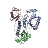
| |||||||||
|---|---|---|---|---|---|---|---|---|---|---|
| 1 |
| |||||||||
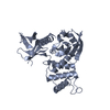
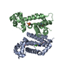
| NMR ensembles |
|
-Components
| #1: Protein | Mass: 21053.768 Da / Num. of mol.: 1 Fragment: N-terminal domain of ataxin-3 sequence database residues 1-182 Source method: isolated from a genetically manipulated source Source: (gene. exp.) Homo sapiens (human) / Gene: ATXN3, ATX3, MJD, MJD1, SCA3 / Production host:  References: UniProt: P54252, Hydrolases; Acting on peptide bonds (peptidases); Cysteine endopeptidases |
|---|---|
| #2: Protein | Mass: 8576.831 Da / Num. of mol.: 2 / Fragment: sequence database residues 318-469 Source method: isolated from a genetically manipulated source Source: (gene. exp.) Homo sapiens (human) / Gene: UBC / Production host: |
-Experimental details
-Experiment
| Experiment | Method: SOLUTION NMR Details: trascription/protein binding, machado-joseph disease protein, ubiquitin-proteasome pathway | ||||||||||||||||||||||||
|---|---|---|---|---|---|---|---|---|---|---|---|---|---|---|---|---|---|---|---|---|---|---|---|---|---|
| NMR experiment |
|
 HSQC
HSQC- Sample preparation
| Details | Contents: 0.1 - 0.3 mM [U-15N] protein, 0.1 -0.3 mM [U-13C; U-15N] protein, 0.1-0.3 mM protein, 0.1-0.3 mM [U-13C; U-15N; U-2H] protein, 90% H2O/10% D2O Solvent system: 90% H2O/10% D2O | ||||||||||||||||||||
|---|---|---|---|---|---|---|---|---|---|---|---|---|---|---|---|---|---|---|---|---|---|
| Sample |
| ||||||||||||||||||||
| Sample conditions | Ionic strength: 0.01 / pH: 6.5 / Pressure: ambient / Temperature: 298 K |
-NMR measurement
| NMR spectrometer |
|
|---|
- Processing
| NMR software |
| ||||||||||||||||
|---|---|---|---|---|---|---|---|---|---|---|---|---|---|---|---|---|---|
| Refinement | Method: torsion angle dynamics / Software ordinal: 1 / Details: refinement in explicit solvent | ||||||||||||||||
| NMR representative | Selection criteria: lowest energy | ||||||||||||||||
| NMR ensemble | Conformer selection criteria: structures with the lowest energy Conformers calculated total number: 100 / Conformers submitted total number: 20 |
