 Movie
Movie Controller

[English] 日本語
 Yorodumi
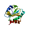
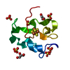
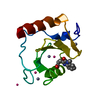
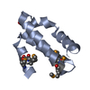
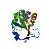
- PDB-5d8v: Ultra-high resolution structure of high-potential iron-sulfur protein -
+ Open data

- Basic information
| Entry | Database: PDB / ID: 5d8v | ||||||
|---|---|---|---|---|---|---|---|
| Title | Ultra-high resolution structure of high-potential iron-sulfur protein | ||||||
 Components Components | High-potential iron-sulfur protein | ||||||
 Keywords Keywords | METAL BINDING PROTEIN / iron-sulfur protein | ||||||
| Function / homology |  Function and homology information Function and homology informationaerobic electron transport chain / 4 iron, 4 sulfur cluster binding / electron transfer activity / metal ion binding Similarity search - Function | ||||||
| Biological species |  Thermochromatium tepidum (bacteria) Thermochromatium tepidum (bacteria) | ||||||
| Method |  X-RAY DIFFRACTION / SYNCHROTRON / MOLECULAR REPLACEMENT / Resolution: 0.48 Å X-RAY DIFFRACTION / SYNCHROTRON / MOLECULAR REPLACEMENT / Resolution: 0.48 Å | ||||||
 Authors Authors | Hirano, Y. / Takeda, K. / Miki, K. | ||||||
 Citation Citation | Journal: Nature / Year: 2016 Title: Charge-density analysis of an iron-sulfur protein at an ultra-high resolution of 0.48 angstrom Authors: Hirano, Y. / Takeda, K. / Miki, K. | ||||||
| History |
|
- Structure visualization
| Structure viewer | Molecule: MolmilJmol/JSmol |
|---|
- Downloads & links
-Download
| PDBx/mmCIF format | 5d8v.cif.gz | 83.8 KB | Display | PDBx/mmCIF format |
|---|---|---|---|---|
| PDB format | pdb5d8v.ent.gz | 64.4 KB | Display | PDB format |
| PDBx/mmJSON format | 5d8v.json.gz | Tree view | PDBx/mmJSON format | |
| Others |  Other downloads Other downloads |
-Validation report
| Arichive directory | https://data.pdbj.org/pub/pdb/validation_reports/d8/5d8vftp://data.pdbj.org/pub/pdb/validation_reports/d8/5d8v | HTTPS FTP |
|---|
-Related structure data
| Related structure data |  3a39S S: Starting model for refinement |
|---|---|
| Similar structure data |


-Links
 PDBj
PDBj- Assembly
| Deposited unit | 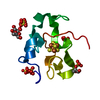
| ||||||||
|---|---|---|---|---|---|---|---|---|---|
| 1 |
| ||||||||
| Unit cell |
|
-Components
| #1: Protein | Mass: 8793.851 Da / Num. of mol.: 1 / Source method: isolated from a natural source / Source: (natural) Thermochromatium tepidum (bacteria) / References: UniProt: P80176 | ||||
|---|---|---|---|---|---|
| #2: Chemical | ChemComp-SF4 /   Mass: 351.640 Da / Num. of mol.: 1 / Source method: obtained synthetically / Formula: Fe4S4 Mass: 351.640 Da / Num. of mol.: 1 / Source method: obtained synthetically / Formula: Fe4S4 | ||||
| #3: Chemical | ChemComp-SO4 /   Mass: 96.063 Da / Num. of mol.: 5 / Source method: obtained synthetically / Formula: SO4 Mass: 96.063 Da / Num. of mol.: 5 / Source method: obtained synthetically / Formula: SO4#4: Chemical |   Mass: 92.094 Da / Num. of mol.: 3 / Source method: obtained synthetically / Formula: C3H8O3 Mass: 92.094 Da / Num. of mol.: 3 / Source method: obtained synthetically / Formula: C3H8O3#5: Water | ChemComp-HOH / |  Mass: 18.015 Da / Num. of mol.: 180 / Source method: isolated from a natural source / Formula: H2O Mass: 18.015 Da / Num. of mol.: 180 / Source method: isolated from a natural source / Formula: H2O |
-Experimental details
-Experiment
| Experiment | Method: X-RAY DIFFRACTION / Number of used crystals: 1 |
|---|
- Sample preparation
| Crystal | Density Matthews: 1.82 Å3/Da / Density % sol: 32.3 % |
|---|---|
| Crystal grow | Temperature: 293 K / Method: vapor diffusion, hanging drop / pH: 4 Details: 1.6 M ammonium sulfate, 0.1 M sodium-citrate, 5 mM DTT |
-Data collection
| Diffraction | Mean temperature: 100 K |
|---|---|
| Diffraction source | Source: SYNCHROTRON / Site: SPring-8  / Beamline: BL41XU / Wavelength: 0.45086 Å / Beamline: BL41XU / Wavelength: 0.45086 Å |
| Detector | Type: RAYONIX MX225HE / Detector: CCD / Date: Feb 23, 2009 / Details: fiber-optic taper |
| Radiation | Monochromator: Double-crystal Si(111) liquid-nitrogen-cooled Protocol: SINGLE WAVELENGTH / Monochromatic (M) / Laue (L): M / Scattering type: x-ray |
| Radiation wavelength | Wavelength: 0.45086 Å / Relative weight: 1 |
| Reflection | Resolution: 0.48→20 Å / Num. obs: 301119 / % possible obs: 96.3 % / Observed criterion σ(I): 0 / Redundancy: 5.4 % / Rmerge(I) obs: 0.056 / Net I/σ(I): 61.1 |
| Reflection shell | Resolution: 0.48→0.5 Å / Redundancy: 3 % / Rmerge(I) obs: 0.339 / Mean I/σ(I) obs: 2.7 / % possible all: 89 |
- Processing
| Software |
| ||||||||||||||||
|---|---|---|---|---|---|---|---|---|---|---|---|---|---|---|---|---|---|
| Refinement | Method to determine structure: MOLECULAR REPLACEMENT Starting model: 3A39 Resolution: 0.48→20 Å / Cross valid method: FREE R-VALUE
| ||||||||||||||||
| Displacement parameters | Biso mean: 6.19 Å2 | ||||||||||||||||
| Refinement step | Cycle: LAST / Resolution: 0.48→20 Å
| ||||||||||||||||
| LS refinement shell | Resolution: 0.48→0.48 Å / Rfactor Rfree: 0.41 / Rfactor Rwork: 0.397 |
