 Movie
Movie Controller


+ Open data

- Basic information
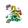
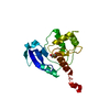
| Entry | Database: PDB / ID: 5d3v | ||||||||||||
|---|---|---|---|---|---|---|---|---|---|---|---|---|---|
| Title | Crystal Structure of the P-Rex1 PH domain with Citrate Bound | ||||||||||||
 Components Components | Phosphatidylinositol 3,4,5-trisphosphate-dependent Rac exchanger 1 protein | ||||||||||||
 Keywords Keywords | LIPID BINDING PROTEIN / pleckstrin homology domain / beta sandwich / phosphatidylinositol-binding | ||||||||||||
| Function / homology |  Function and homology information Function and homology informationregulation of dendrite development / regulation of actin filament polymerization / neutrophil activation / regulation of small GTPase mediated signal transduction / negative regulation of TOR signaling / RHOB GTPase cycle / superoxide metabolic process / NRAGE signals death through JNK / RHOJ GTPase cycle / RHOC GTPase cycle ...regulation of dendrite development / regulation of actin filament polymerization / neutrophil activation / regulation of small GTPase mediated signal transduction / negative regulation of TOR signaling / RHOB GTPase cycle / superoxide metabolic process / NRAGE signals death through JNK / RHOJ GTPase cycle / RHOC GTPase cycle / RHOQ GTPase cycle / CDC42 GTPase cycle / T cell differentiation / RHOG GTPase cycle / RAC3 GTPase cycle / RHOA GTPase cycle / RAC2 GTPase cycle / protein serine/threonine kinase inhibitor activity / RAC1 GTPase cycle / actin filament polymerization / neutrophil chemotaxis / positive regulation of substrate adhesion-dependent cell spreading / guanyl-nucleotide exchange factor activity / GTPase activator activity / dendritic shaft / phospholipid binding / G alpha (12/13) signalling events / growth cone / intracellular signal transduction / positive regulation of cell migration / G protein-coupled receptor signaling pathway / perinuclear region of cytoplasm / enzyme binding / plasma membrane / cytosol Similarity search - Function | ||||||||||||
| Biological species |  Homo sapiens (human) Homo sapiens (human) | ||||||||||||
| Method |  X-RAY DIFFRACTION / SYNCHROTRON / MOLECULAR REPLACEMENT / molecular replacement / Resolution: 1.852 Å X-RAY DIFFRACTION / SYNCHROTRON / MOLECULAR REPLACEMENT / molecular replacement / Resolution: 1.852 Å | ||||||||||||
 Authors Authors | Cash, J.N. / Tesmer, J.J.G. | ||||||||||||
| Funding support |  United States, 3items United States, 3items
| ||||||||||||
 Citation Citation | Journal: Structure / Year: 2016 Title: Structural and Biochemical Characterization of the Catalytic Core of the Metastatic Factor P-Rex1 and Its Regulation by PtdIns(3,4,5)P3. Authors: Cash, J.N. / Davis, E.M. / Tesmer, J.J. | ||||||||||||
| History |
|
- Structure visualization
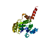
| Structure viewer | Molecule: MolmilJmol/JSmol |
|---|
- Downloads & links
-Download
| PDBx/mmCIF format | 5d3v.cif.gz | 79.5 KB | Display | PDBx/mmCIF format |
|---|---|---|---|---|
| PDB format | pdb5d3v.ent.gz | 57.3 KB | Display | PDB format |
| PDBx/mmJSON format | 5d3v.json.gz | Tree view | PDBx/mmJSON format | |
| Others |  Other downloads Other downloads |
-Validation report
| Arichive directory | https://data.pdbj.org/pub/pdb/validation_reports/d3/5d3vftp://data.pdbj.org/pub/pdb/validation_reports/d3/5d3v | HTTPS FTP |
|---|
-Related structure data
| Related structure data | 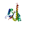 5d27C 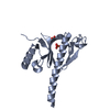 5d3wC 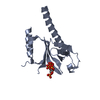 5d3xC 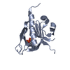 5d3yC 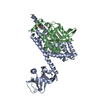 5fi0C 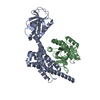 5fi1C 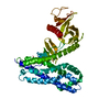 2pz1S C: citing same article ( S: Starting model for refinement |
|---|---|
| Similar structure data |


-Links
 PDBj
PDBj


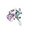
- Assembly
| Deposited unit | 
| ||||||||
|---|---|---|---|---|---|---|---|---|---|
| 1 | 
| ||||||||
| 2 | 
| ||||||||
| Unit cell |
|
-Components
| #1: Protein | Mass: 19286.953 Da / Num. of mol.: 2 / Fragment: unp residues 245-408 Source method: isolated from a genetically manipulated source Details: pleckstrin homology domain / Source: (gene. exp.) Homo sapiens (human) / Gene: PREX1, KIAA1415 / Plasmid: pMALc2H10T / Production host:  #2: Chemical | ChemComp-FLC / |   Mass: 189.100 Da / Num. of mol.: 1 / Source method: obtained synthetically / Formula: C6H5O7 Mass: 189.100 Da / Num. of mol.: 1 / Source method: obtained synthetically / Formula: C6H5O7#3: Water | ChemComp-HOH / |  Mass: 18.015 Da / Num. of mol.: 222 / Source method: isolated from a natural source / Formula: H2O Mass: 18.015 Da / Num. of mol.: 222 / Source method: isolated from a natural source / Formula: H2O |
|---|
-Experimental details
-Experiment
| Experiment | Method: X-RAY DIFFRACTION / Number of used crystals: 1 |
|---|
- Sample preparation
| Crystal | Density Matthews: 2.09 Å3/Da / Density % sol: 41.29 % |
|---|---|
| Crystal grow | Temperature: 294 K / Method: vapor diffusion, hanging drop / pH: 5 / Details: Sodium citrate, PEG 8000 |
-Data collection
| Diffraction | Mean temperature: 110 K | ||||||||||||||||||||||||||||||||||||||||||||||||||||||||||||||||||||||||||||||||||||||||||||||||||||||||||||||||||||||||||||||
|---|---|---|---|---|---|---|---|---|---|---|---|---|---|---|---|---|---|---|---|---|---|---|---|---|---|---|---|---|---|---|---|---|---|---|---|---|---|---|---|---|---|---|---|---|---|---|---|---|---|---|---|---|---|---|---|---|---|---|---|---|---|---|---|---|---|---|---|---|---|---|---|---|---|---|---|---|---|---|---|---|---|---|---|---|---|---|---|---|---|---|---|---|---|---|---|---|---|---|---|---|---|---|---|---|---|---|---|---|---|---|---|---|---|---|---|---|---|---|---|---|---|---|---|---|---|---|---|
| Diffraction source | Source: SYNCHROTRON / Site: APS / Beamline: 21-ID-D / Wavelength: 1.078 Å | ||||||||||||||||||||||||||||||||||||||||||||||||||||||||||||||||||||||||||||||||||||||||||||||||||||||||||||||||||||||||||||||
| Detector | Type: MARMOSAIC 300 mm CCD / Detector: CCD / Date: Feb 5, 2014 | ||||||||||||||||||||||||||||||||||||||||||||||||||||||||||||||||||||||||||||||||||||||||||||||||||||||||||||||||||||||||||||||
| Radiation | Protocol: SINGLE WAVELENGTH / Monochromatic (M) / Laue (L): M / Scattering type: x-ray | ||||||||||||||||||||||||||||||||||||||||||||||||||||||||||||||||||||||||||||||||||||||||||||||||||||||||||||||||||||||||||||||
| Radiation wavelength | Wavelength: 1.078 Å / Relative weight: 1 | ||||||||||||||||||||||||||||||||||||||||||||||||||||||||||||||||||||||||||||||||||||||||||||||||||||||||||||||||||||||||||||||
| Reflection | Resolution: 1.85→50 Å / Num. obs: 27997 / % possible obs: 99.5 % / Redundancy: 4 % / Biso Wilson estimate: 22.58 Å2 / Rmerge(I) obs: 0.054 / Χ2: 1.177 / Net I/av σ(I): 25.811 / Net I/σ(I): 11.9 / Num. measured all: 111287 | ||||||||||||||||||||||||||||||||||||||||||||||||||||||||||||||||||||||||||||||||||||||||||||||||||||||||||||||||||||||||||||||
| Reflection shell | Diffraction-ID: 1 / Rejects: _
|
-Phasing
| Phasing | Method: molecular replacement |
|---|
- Processing
| Software |
| |||||||||||||||||||||||||||||||||||||||||||||||||||||||||||||||||||||||||||||
|---|---|---|---|---|---|---|---|---|---|---|---|---|---|---|---|---|---|---|---|---|---|---|---|---|---|---|---|---|---|---|---|---|---|---|---|---|---|---|---|---|---|---|---|---|---|---|---|---|---|---|---|---|---|---|---|---|---|---|---|---|---|---|---|---|---|---|---|---|---|---|---|---|---|---|---|---|---|---|
| Refinement | Method to determine structure: MOLECULAR REPLACEMENT Starting model: 2PZ1 Resolution: 1.852→36.192 Å / SU ML: 0.21 / Cross valid method: THROUGHOUT / σ(F): 1.36 / Phase error: 21.19 / Stereochemistry target values: ML
| |||||||||||||||||||||||||||||||||||||||||||||||||||||||||||||||||||||||||||||
| Solvent computation | Shrinkage radii: 0.9 Å / VDW probe radii: 1.11 Å / Solvent model: FLAT BULK SOLVENT MODEL | |||||||||||||||||||||||||||||||||||||||||||||||||||||||||||||||||||||||||||||
| Displacement parameters | Biso max: 101.14 Å2 / Biso mean: 33.8325 Å2 / Biso min: 9.05 Å2 | |||||||||||||||||||||||||||||||||||||||||||||||||||||||||||||||||||||||||||||
| Refinement step | Cycle: final / Resolution: 1.852→36.192 Å
| |||||||||||||||||||||||||||||||||||||||||||||||||||||||||||||||||||||||||||||
| Refine LS restraints |
| |||||||||||||||||||||||||||||||||||||||||||||||||||||||||||||||||||||||||||||
| LS refinement shell | Refine-ID: X-RAY DIFFRACTION / Total num. of bins used: 10
|
