 Movie
Movie Controller

[English] 日本語
 Yorodumi
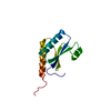
- PDB-5d22: Structure of ovine granulocyte-macrophage colony-stimulating factor -
+ Open data

- Basic information
| Entry | Database: PDB / ID: 5d22 | ||||||
|---|---|---|---|---|---|---|---|
| Title | Structure of ovine granulocyte-macrophage colony-stimulating factor | ||||||
 Components Components | Granulocyte-macrophage colony-stimulating factor | ||||||
 Keywords Keywords | CYTOKINE / Signaling Protein / recombinant proteins / cytokine immunology | ||||||
| Function / homology |  Function and homology information Function and homology informationgranulocyte macrophage colony-stimulating factor receptor binding / neutrophil differentiation / positive regulation of interleukin-23 production / granulocyte macrophage colony-stimulating factor receptor complex / granulocyte-macrophage colony-stimulating factor signaling pathway / positive regulation of leukocyte proliferation / myeloid dendritic cell differentiation / cellular response to granulocyte macrophage colony-stimulating factor stimulus / positive regulation of macrophage derived foam cell differentiation / positive regulation of podosome assembly ...granulocyte macrophage colony-stimulating factor receptor binding / neutrophil differentiation / positive regulation of interleukin-23 production / granulocyte macrophage colony-stimulating factor receptor complex / granulocyte-macrophage colony-stimulating factor signaling pathway / positive regulation of leukocyte proliferation / myeloid dendritic cell differentiation / cellular response to granulocyte macrophage colony-stimulating factor stimulus / positive regulation of macrophage derived foam cell differentiation / positive regulation of podosome assembly / monocyte differentiation / macrophage differentiation / cell surface receptor signaling pathway via JAK-STAT / negative regulation of extrinsic apoptotic signaling pathway in absence of ligand / embryonic placenta development / cytokine activity / growth factor activity / cell population proliferation / immune response / negative regulation of DNA-templated transcription / intracellular membrane-bounded organelle / extracellular space Similarity search - Function | ||||||
| Biological species |  | ||||||
| Method |  X-RAY DIFFRACTION / SYNCHROTRON / MOLECULAR REPLACEMENT / Resolution: 1.994 Å X-RAY DIFFRACTION / SYNCHROTRON / MOLECULAR REPLACEMENT / Resolution: 1.994 Å | ||||||
 Authors Authors | Felix, J. / Savvides, S.N. | ||||||
 Citation Citation | Journal: Nat Commun / Year: 2016 Title: Structural basis of GM-CSF and IL-2 sequestration by the viral decoy receptor GIF. Authors: Jan Felix / Eaazhisai Kandiah / Steven De Munck / Yehudi Bloch / Gydo C P van Zundert / Kris Pauwels / Ann Dansercoer / Katka Novanska / Randy J Read / Alexandre M J J Bonvin / Bjorn ...Authors: Jan Felix / Eaazhisai Kandiah / Steven De Munck / Yehudi Bloch / Gydo C P van Zundert / Kris Pauwels / Ann Dansercoer / Katka Novanska / Randy J Read / Alexandre M J J Bonvin / Bjorn Vergauwen / Kenneth Verstraete / Irina Gutsche / Savvas N Savvides /      Abstract: Subversion of the host immune system by viruses is often mediated by molecular decoys that sequester host proteins pivotal to mounting effective immune responses. The widespread mammalian pathogen ...Subversion of the host immune system by viruses is often mediated by molecular decoys that sequester host proteins pivotal to mounting effective immune responses. The widespread mammalian pathogen parapox Orf virus deploys GIF, a member of the poxvirus immune evasion superfamily, to antagonize GM-CSF (granulocyte macrophage colony-stimulating factor) and IL-2 (interleukin-2), two pleiotropic cytokines of the mammalian immune system. However, structural and mechanistic insights into the unprecedented functional duality of GIF have remained elusive. Here we reveal that GIF employs a dimeric binding platform that sequesters two copies of its target cytokines with high affinity and slow dissociation kinetics to yield distinct complexes featuring mutually exclusive interaction footprints. We illustrate how GIF serves as a competitive decoy receptor by leveraging binding hotspots underlying the cognate receptor interactions of GM-CSF and IL-2, without sharing any structural similarity with the cytokine receptors. Our findings contribute to the tracing of novel molecular mimicry mechanisms employed by pathogenic viruses. | ||||||
| History |
|
- Structure visualization
| Structure viewer | Molecule: MolmilJmol/JSmol |
|---|
- Downloads & links
-Download
| PDBx/mmCIF format | 5d22.cif.gz | 111 KB | Display | PDBx/mmCIF format |
|---|---|---|---|---|
| PDB format | pdb5d22.ent.gz | 84.9 KB | Display | PDB format |
| PDBx/mmJSON format | 5d22.json.gz | Tree view | PDBx/mmJSON format | |
| Others |  Other downloads Other downloads |
-Validation report
| Summary document | 5d22_validation.pdf.gz | 452.1 KB | Display | wwPDB validaton report |
|---|---|---|---|---|
| Full document | 5d22_full_validation.pdf.gz | 454 KB | Display | |
| Data in XML | 5d22_validation.xml.gz | 12.2 KB | Display | |
| Data in CIF | 5d22_validation.cif.gz | 16.4 KB | Display | |
| Arichive directory | https://data.pdbj.org/pub/pdb/validation_reports/d2/5d22ftp://data.pdbj.org/pub/pdb/validation_reports/d2/5d22 | HTTPS FTP |
-Related structure data
| Related structure data |  4060C  5d28C  2gmfS S: Starting model for refinement C: citing same article ( |
|---|---|
| Similar structure data |


-Links
 PDBj
PDBj


- Assembly
| Deposited unit | 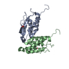
| ||||||||
|---|---|---|---|---|---|---|---|---|---|
| 1 | 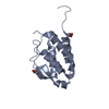
| ||||||||
| 2 | 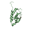
| ||||||||
| Unit cell |
|
-Components
| #1: Protein | Mass: 14426.386 Da / Num. of mol.: 2 Source method: isolated from a genetically manipulated source Source: (gene. exp.)  #2: Chemical |   Mass: 59.044 Da / Num. of mol.: 2 / Source method: obtained synthetically / Formula: C2H3O2 Mass: 59.044 Da / Num. of mol.: 2 / Source method: obtained synthetically / Formula: C2H3O2#3: Chemical | ChemComp-EDO / |   Mass: 62.068 Da / Num. of mol.: 1 / Source method: obtained synthetically / Formula: C2H6O2 Mass: 62.068 Da / Num. of mol.: 1 / Source method: obtained synthetically / Formula: C2H6O2#4: Water | ChemComp-HOH / |  Mass: 18.015 Da / Num. of mol.: 121 / Source method: isolated from a natural source / Formula: H2O Mass: 18.015 Da / Num. of mol.: 121 / Source method: isolated from a natural source / Formula: H2OHas protein modification | Y | |
|---|
-Experimental details
-Experiment
| Experiment | Method: X-RAY DIFFRACTION |
|---|
- Sample preparation
| Crystal | Density Matthews: 2.42 Å3/Da / Density % sol: 49.2 % |
|---|---|
| Crystal grow | Temperature: 293 K / Method: vapor diffusion, sitting drop / pH: 8.5 Details: 0.2 M sodium acetate trihydrate 0.1 M TRIS hydrochloride pH 8.5 30% w/v PEG 4000 |
-Data collection
| Diffraction | Mean temperature: 100 K |
|---|---|
| Diffraction source | Source: SYNCHROTRON / Site: PETRA III, EMBL c/o DESY  / Beamline: P14 (MX2) / Wavelength: 1.239 Å / Beamline: P14 (MX2) / Wavelength: 1.239 Å |
| Detector | Type: DECTRIS PILATUS 6M / Detector: PIXEL / Date: Nov 16, 2012 |
| Radiation | Protocol: SINGLE WAVELENGTH / Monochromatic (M) / Laue (L): M / Scattering type: x-ray |
| Radiation wavelength | Wavelength: 1.239 Å / Relative weight: 1 |
| Reflection | Resolution: 1.99→38.51 Å / Num. all: 18842 / Num. obs: 18252 / % possible obs: 96.9 % / Redundancy: 3.28 % / Rsym value: 0.129 / Net I/σ(I): 8.17 |
| Reflection shell | Resolution: 1.99→2.11 Å / Redundancy: 3.14 % / Mean I/σ(I) obs: 2.12 / Rsym value: 0.732 / % possible all: 94 |
- Processing
| Software |
| |||||||||||||||||||||||||||||||||||||||||||||||||
|---|---|---|---|---|---|---|---|---|---|---|---|---|---|---|---|---|---|---|---|---|---|---|---|---|---|---|---|---|---|---|---|---|---|---|---|---|---|---|---|---|---|---|---|---|---|---|---|---|---|---|
| Refinement | Method to determine structure: MOLECULAR REPLACEMENT Starting model: 2GMF Resolution: 1.994→38.51 Å / SU ML: 0.27 / Cross valid method: FREE R-VALUE / σ(F): 1.99 / Phase error: 26.09 / Stereochemistry target values: ML
| |||||||||||||||||||||||||||||||||||||||||||||||||
| Solvent computation | Shrinkage radii: 0.9 Å / VDW probe radii: 1.11 Å / Solvent model: FLAT BULK SOLVENT MODEL | |||||||||||||||||||||||||||||||||||||||||||||||||
| Refinement step | Cycle: LAST / Resolution: 1.994→38.51 Å
| |||||||||||||||||||||||||||||||||||||||||||||||||
| Refine LS restraints |
| |||||||||||||||||||||||||||||||||||||||||||||||||
| LS refinement shell |
| |||||||||||||||||||||||||||||||||||||||||||||||||
| Refinement TLS params. | Method: refined / Origin x: 3.6389 Å / Origin y: -18.0912 Å / Origin z: -9.3264 Å
| |||||||||||||||||||||||||||||||||||||||||||||||||
| Refinement TLS group | Selection details: all |
